

Unlocking Benzosampangine's Potential: A Computational Approach to Investigating, Its Role as a PD-L1 Inhibitor in Tumor Immune Evasion via Molecular Docking, Dynamic Simulation, and ADMET Profiling
- PMID: 39564188
- PMCID: PMC11574905
- DOI: 10.1177/11779322241298591
Unlocking Benzosampangine's Potential: A Computational Approach to Investigating, Its Role as a PD-L1 Inhibitor in Tumor Immune Evasion via Molecular Docking, Dynamic Simulation, and ADMET Profiling
Abstract
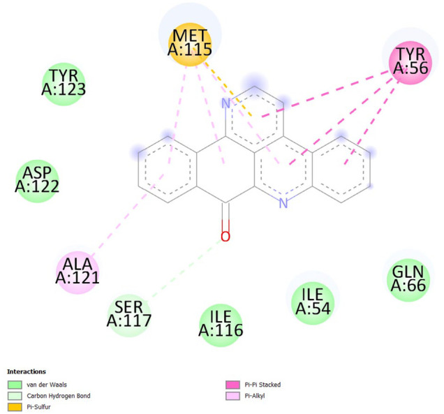
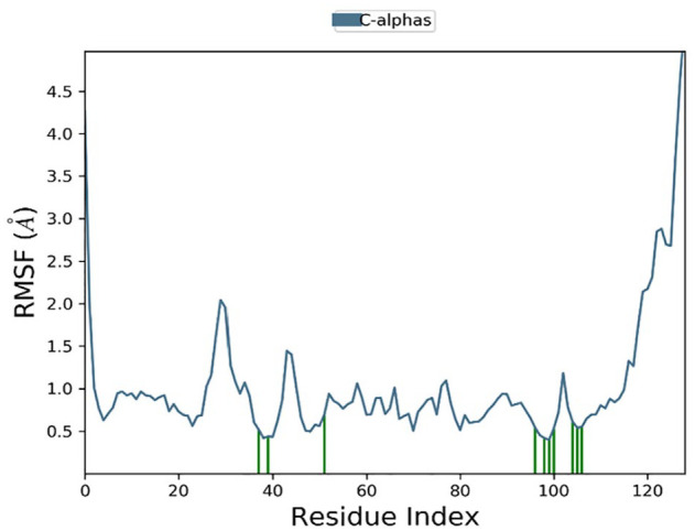
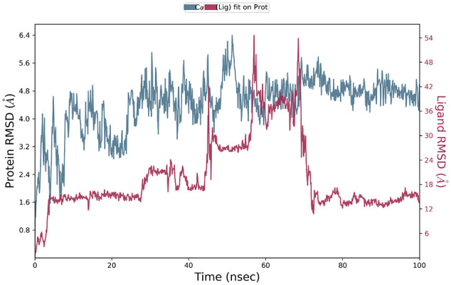
The interaction between programmed cell death protein 1 (PD-1) and its ligand PD-L1 plays a crucial role in tumor immune evasion, presenting a critical target for cancer immunotherapy. Despite being effective, current monoclonal antibodies present some drawbacks such as high costs, toxicity, and resistance development. Therefore, the development of small-molecule inhibitors is necessary, especially those derived from natural sources. In this study, benzosampangine is predicted as a promising PD-L1 inhibitor, with potential applications in cancer immunotherapy. Utilizing the high-resolution crystal structure of human PD-L1 (PDB ID: 5O45), we screened 511 natural compounds, identifying benzosampangine as a top candidate with exceptional inhibitory properties. Molecular docking predicted that benzosampangine exhibits a strong binding affinity for PD-L1 (-9.4 kcal/mol) compared with established controls such as CA-170 (-6.5 kcal/mol), BMS-202 (-8.6 kcal/mol), and pyrvinium (-8.9 kcal/mol). The compound's predicted binding efficacy is highlighted by robust interactions with key amino acids (ILE54, TYR56, GLN66, MET115, ILE116, SER117, ALA121, ASP122) within the active site, notably forming 3 Pi-sulfur interactions with MET115-an interaction absents in control inhibitors. In addition, ADMET profiling suggests that over the control molecules, benzosampangine has several key advantages, including favorable solubility, permeability, metabolic stability, and low toxicity, while adhering to Lipinski's rule of five. Molecular dynamic simulations predict the stability of the benzosampangine-PD-L1 complex, reinforcing its potential to sustain inhibition of the PD-1/PD-L1 pathway. MMGBSA analysis calculated a binding free energy (ΔGbind) of -39.39 kcal/mol for the benzosampangine-PD-L1 complex, with significant contributions from Coulombic, lipophilic, and Van der Waals interactions, validating the predicted docking results. This study investigates in silico benzosampangine, predicting its better molecular interactions and pharmacokinetic profile compared with several already known PD-L1 inhibitors.
Keywords: PD-1/PD-L1 inhibitors; benzosampangine; cancer; in silico study; molecular docking; molecular dynamic; small molecules.
© The Author(s) 2024.
Conflict of interest statement
The author(s) declared no potential conflicts of interest with respect to the research, authorship, and/or publication of this article.
Figures


Similar articles
-
Molecular Mechanism of Small-Molecule Inhibitors in Blocking the PD-1/PD-L1 Pathway through PD-L1 Dimerization.Int J Mol Sci. 2021 Apr 30;22(9):4766. doi: 10.3390/ijms22094766. Int J Mol Sci. 2021. PMID: 33946261 Free PMC article.
-
A computational chemistry-based approach to optimizing PD-1/PD-L1 inhibitors.Front Chem. 2025 Jan 14;12:1533541. doi: 10.3389/fchem.2024.1533541. eCollection 2024. Front Chem. 2025. PMID: 39877119 Free PMC article.
-
Is the Triggering of PD-L1 Dimerization a Potential Mechanism for Food-Derived Small Molecules in Cancer Immunotherapy? A Study by Molecular Dynamics.Int J Mol Sci. 2023 Jan 11;24(2):1413. doi: 10.3390/ijms24021413. Int J Mol Sci. 2023. PMID: 36674929 Free PMC article.
-
Biphenyl-based small molecule inhibitors: Novel cancer immunotherapeutic agents targeting PD-1/PD-L1 interaction.Bioorg Med Chem. 2022 Nov 1;73:117001. doi: 10.1016/j.bmc.2022.117001. Epub 2022 Sep 13. Bioorg Med Chem. 2022. PMID: 36126447 Review.
-
Peptide-based and small synthetic molecule inhibitors on PD-1/PD-L1 pathway: A new choice for immunotherapy?Eur J Med Chem. 2019 Jan 1;161:378-398. doi: 10.1016/j.ejmech.2018.10.044. Epub 2018 Oct 19. Eur J Med Chem. 2019. PMID: 30384043 Review.
References
-
- Catros-Quemener V, Bouet et NoëlleGenetet F. Immunité thérapies cellulaires du cancer. Med Sci (Paris). 2003;19:43-53. - PubMed
-
- Walker PR, Prins RM, Dietrich P-Y, Liau LM. Harnessing T-cell immunity to target brain tumors. In: Van Meir EG, ed. CNS Cancer. Humana Press; 2009:1165-1217. doi:10.1007/978-1-60327-553-8_48 - DOI
LinkOut - more resources
Full Text Sources
Research Materials
Miscellaneous
