. 2024 Dec;291(24):5506-5522.
doi: 10.1111/febs.17328. Epub 2024 Nov 20.

Interaction with the cysteine-free protein HAX1 expands the substrate specificity and function of MIA40 beyond protein oxidation

Affiliations

Interaction with the cysteine-free protein HAX1 expands the substrate specificity and function of MIA40 beyond protein oxidation

Robin Alexander Rothemann et al. FEBS J. 2024 Dec.

Abstract

The mitochondrial disulphide relay machinery is essential for the import and oxidative folding of many proteins in the mitochondrial intermembrane space. Its core component, the import receptor MIA40 (also CHCHD4), serves as an oxidoreductase but also as a chaperone holdase, which initially interacts with its substrates non-covalently before introducing disulphide bonds for folding and retaining proteins in the intermembrane space. Interactome studies have identified diverse substrates of MIA40, among them the intrinsically disordered HCLS1-associated protein X-1 (HAX1). Interestingly, this protein does not contain cysteines, raising the question of how and to what end HAX1 can interact with MIA40. Here, we demonstrate that MIA40 non-covalently interacts with HAX1 independent of its redox-active cysteines. While HAX1 import is driven by its weak mitochondrial targeting sequence, its subsequent transient interaction with MIA40 stabilizes the protein in the intermembrane space. HAX1 solely depends on the holdase activity of MIA40, and the absence of MIA40 results in the aggregation, degradation and loss of HAX1. Collectively, our study introduces HAX1 as the first endogenous MIA40 substrate without cysteines and demonstrates the diverse functions of this highly conserved oxidoreductase and import receptor.

Keywords: HAX1; IMS; MIA40; mitochondria; mitochondrial disulphide relay.


Conflict of interest statement

The authors declare no conflicts of interest.

Figures

Fig. 1
Fig. 1
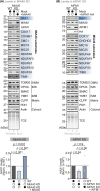
HAX1 interacts with MIA40 in a noncovalent manner. (A) HAX1 is enriched in different MIA40 interactomes. Two independent interactome studies of MIA40 identified HAX1 as significantly enriched with MIA40, similar to known MIA40 substrates [13, 34]. However, interaction could only be detected if the immunoprecipitation of MIA40 had been performed after native cell lysis. (B) Domain layout of HAX1. HAX1 is devoid of any cysteine residues. Most of its N‐terminal amino acids do not, according to TargetP, serve as MTS as they also contain negatively charged amino acid residues. However, a sliding TargetP score prediction (iMTS score) indicates a certain propensity of this region to serve as MTS. Moreover, HAX1 contains a conserved amphipathic helix (between residues 81 and 95) that might facilitate MIA40 interaction. MTS, mitochondrial targeting signal; aa, amino acid; structure modelled with Alphafold 2.0. (C) HAX1 localizes to the IMS in a split GFP assay. GFP can be split into two non‐fluorescent parts, a larger part (GFP1‐10) and a smaller part (GFP11). If these parts come together in the same compartment, they can self‐reassemble to reconstitute a fluorescent GFP. GFP1‐10 was equipped either with an MTS for the matrix (Su9MTS) or for the IMS (Sco2MTS) or expressed without targeting information in the cytosol. GFP11 was C‐terminally fused to full‐length HAX1. Reconstitution of GFP fluorescence was successful only for Sco2‐GFP1‐10 and HAX1‐GFP11, indicating that HAX1 is present in the IMS. For matrix‐localized GFP1‐10, a weak background fluorescence was observable. Mitotracker signal served as positive control for mitochondria. Magenta, mitotracker signal; Cyan, GFP signal. Bar corresponds to 10 μm. N = 2 replicates. (D) Endogenous HAX1 localizes to mitochondria in different cell lines. The indicated cells were fixated, permeabilized, and stained using a primary antibody against HAX1 or mitotracker. Cells were analysed by fluorescence microscopy. Mitotracker signal served as positive control for mitochondria. Magenta, mitotracker signal; Cyan, HAX1 signal. A lilac/whitish colour indicates signal overlap. Bar corresponds to 10 μm. N = 2 replicates. (E) Digitonin fractionation to detect the localization of endogenous HAX1. HEK293 cells were incubated with increasing amounts of digitonin, leading to a step‐wise solubilization of cellular membranes. After centrifugation, the supernatant and pellet were analysed by SDS‐PAGE and immunoblot. PSMC4, CPOX and PDH served as controls for cytosol, IMS and matrix, respectively. HAX1 behaved very similar to the IMS control CPOX. 2,2,2‐Trichloroethanol (TCE) incorporated into gels before polymerization provided fluorescent visible detection of proteins as loading control. N = 2 replicates. (F) Endogenous HAX1 in HEK293 cells does not contain cysteine residues. To test for the presence of cysteines in endogenous HAX1, cells were lysed and treated with the strong reductant TCEP. Lysates were either incubated with the maleimide mmPEG24 or left untreated. mmPEG24 modifies free thiols, resulting in a shift per labelled cysteine residue to a higher molecular mass. Lysates were analysed by SDS‐PAGE and immunoblotting. The change in SDS‐PAGE migration behaviour of MIA40 with its seven cysteines served as positive control. Asterisk, background band. Black and white circles represent the indicated treatment and not‐treated samples, respectively. N = 2 replicates. (G) HAX1 coprecipitates with MIA40 in native but not denaturing immunoprecipitation. MIA40‐HA was immunoprecipitated (IP) under native (left panel) and denaturing (right panel) conditions after stopping thiol‐disulphide exchange reactions by NEM incubation. Precipitates were tested for HA (MIA40) HAX1, and the known MIA40 interaction partners NDUFS5 and COX6B1 by reducing SDS‐PAGE and immunoblotting. 1% of the total lysate was loaded as input control. TCE staining served as loading control. Black and white circles represent the indicated treatment and not‐treated samples. N = 3 replicates. (H) HAX1 interacts in vitro with purified recombinant MIA40 irrespective of the presence of the redox‐active motif of MIA40, but displays a reduced interaction with the chaperone‐inactive MIA40 variant. GST‐tagged variants of MIA40 (wild‐type, WT, redox‐inactive C4, 53, 55S and chaperone‐inactive F68E) were purified and bound to beads. The beads were incubated with in vitro translated radioactive HAX1. Subsequently, beads were washed and analysed for bound HAX1 by SDS‐PAGE and autoradiography. Incubation with empty beads served as control (lanes 3, 6, 10 and 13). 0.1% of the total lysate was loaded as input control (lanes 2 and 9). WT MIA40 (lanes 4 and 11) interacts with HAX1 as does the MIA40 C4, 53, 53S variant (lane 7). The MIA40 F68E variant exhibits lower binding to HAX1 (lane 14). TCE staining served as loading control. For quantification, the co‐precipitated HAX1 levels were normalized to the levels precipitated by WT MIA40 and in addition normalized to the according MIA40 signal in the TCE staining. Black and white circles represent the indicated treatment and not‐treated samples. N = 3 (WT), 1 (SPS) and 2 (F68E) replicates.
Fig. 2
Fig. 2
HAX1 import does not critically depend on MIA40. (A) In vitro import of HAX1 is only slightly impaired in mitochondria isolated from MIA40 knockdown (MIA40 KD) compared to wild‐type (WT) cells. In vitro translated radioactive HAX1 was incubated with mitochondria isolated from HEK293 cells (wild‐type, WT and MIA40 knockdown, MIA40 KD). Non‐imported proteins were removed by treatment with Proteinase K (PK). An import reaction was performed into mitochondria treated with CCCP to dissipate the mitochondrial membrane potential. Imported proteins were analysed by reducing SDS‐PAGE and autoradiography. Signals were quantified using ImageQuantTL, and the amount of imported protein was normalized to the maximal signal of the import reaction and plotted. HAX1 can be imported into mitochondria and relies on the mitochondrial membrane potential for import. Depletion of MIA40 has only a minor effect compared to the known MIA substrate COX19. Import of the matrix protein SOD2 served as control for a MIA40‐independent protein. N = 4 replicates; error bars indicate SD. (B) Submitochondrial fractionation after HAX1 import into isolated mitochondria. As (A), except that after import, mitochondria were fractionated by either treating them with an isotonic, a hypotonic (OMM opens) or a Triton X100 (TX‐100, mitochondria are completely lysed)‐containing buffer and then incubating them in the presence or absence of Proteinase K (PK). Marker proteins for different mitochondrial compartments served as fractionation controls. The left panel shows the PK‐treated samples of the right panel for comparison, indicating that newly imported HAX1 is only protected from PK treatment in intact mitochondria but not in mitochondria lacking the OMM, thereby placing it in the IMS. N = 1 replicate. (C) HAX1 import into isolated mitochondria depends on its N‐terminal 26 amino acids. As (A), except that HAX1 wild‐type and a HAX1 variant lacking the N‐terminal, 26 amino acid residues that are predicted to serve as MTS were imported into mitochondria isolated from wild‐type HEK293 cells. N = 4 replicates; error bars indicate SD. (D) Import of a HAX1 mutant lacking its N‐terminal 26 amino acids is only slightly dependent on MIA40. As (A), except that the HAX1 variant lacking the N‐terminal, 26 amino acid residues are imported into mitochondria isolated from wild‐type or MIA40 KD HEK293 cells. N = 4 replicates; error bars indicate SD. Black and white circles represent the indicated treatment and not‐treated samples.
Fig. 3
Fig. 3
HAX1 levels are decreased in cells lacking MIA40 or AIFM1. (A, B) Levels of HAX1 in cells lacking MIA40 (MIA40 KD) (A) or AIFM1 (AIFM1 KO) (B) are decreased. Protein levels in HEK293 cell lines depleted of MIA40 (A) or AIFM1 (B) or overexpressing both proteins were compared to protein levels in wild‐type HEK293 cells. Lysates were analysed by reducing SDS‐PAGE and immunoblotting. Signals were quantified using ImageLab, and the amount of protein was plotted. N = 2–6 replicates; white circles indicate the quantification of individual experiments. TCE staining served as loading control. Black and white circles represent the indicated treatment and not‐treated samples. N = 5 (WT, MIA40 KD), 2 (MIA40 KD + MIA40), N = 6 (WT, AIFM1 KO, AIFM1 KO + AIFM1) replicates.
Fig. 4
Fig. 4
MIA40 functions as a chaperone holdase and stabilizes HAX1. (A) MIA40 suppresses aggregation of citrate synthase (CS). Purified wild‐type MIA40 was added in 10‐, 20‐ and 40‐fold molar excess to chemically denatured citrate synthase, and light scattering was measured. Light scattering in the absence of MIA40 served as a negative control. As a positive control, a 10‐fold excess of chaperone‐active Get3 was added to citrate synthase. N = 2 replicates. (B) MIA40 suppresses aggregation of HAX1. Purified recombinant MIA40 variants (wild‐type, WT and chaperone‐inactive F68E) were incubated with in vitro translated radioactive HAX1 for 1 h. Then, the sample was separated into soluble (S) and insoluble (P) components by ultracentrifugation and analysed by SDS‐PAGE and autoradiography. While addition of WT MIA40 led to increased amounts of HAX1 in the soluble fraction, the holdase‐inactive F68E variant did not affect the P/S ratio. Signals were quantified using ImageQuantTL. The ratio in absence of MIA40 was set to 1.0. T, total; TSP, total‐supernatant‐pellet assay. Black and white circles represent the indicated treatment and not‐treated samples. N = 4 replicates. (C) Loss of MIA40 results in lower levels of soluble HAX1 in mitochondria. Mitochondria were isolated from wild‐type (WT) cells or cells depleted of MIA40 (MIA40 KD). Mitochondria were opened and separated into soluble (S) and insoluble (P) components using a carbonate extraction‐ultracentrifugation approach and analysed by SDS‐PAGE and autoradiography. Absence of MIA40 decreased the amounts of soluble HAX1. T, total; TSP, total‐supernatant‐pellet assay. Black and white circles represent the indicated treatment and not‐treated samples. N = 2 replicates. (D) The HAX1–MIA40 interaction is transient in an emetine chase‐coupled immunoprecipitation assay. HEK293 cells stably expressing MIA40‐HA were treated for the indicated times with the translation inhibitor emetine. Then, cells were lysed under native conditions (Triton X‐100), and MIA40‐HA was precipitated. Precipitates were analysed by reducing SDS–PAGE and immunoblotting. MIA40 and HAX1 interact transiently with each other comparable to the MIA40 substrate NDUFS5. Conversely, the MIA40‐AIFM1 interaction is stable. Asterisk, background band. TCE staining served as loading control. N = 2 replicates. (E) Model. HAX1 import into the IMS is mainly driven by its weak MTS. The intrinsically disordered protein is stabilized by MIA40 via a transient and non‐covalent interaction. In the absence of MIA40, HAX1 is unstable and prone to degradation by proteases.

References

    1. Hung V, Zou P, Rhee HW, Udeshi ND, Cracan V, Svinkina T, Carr SA, Mootha VK & Ting AY (2014) Proteomic mapping of the human mitochondrial intermembrane space in live cells via ratiometric APEX tagging. Mol Cell 55, 332–341. - PMC - PubMed
    1. Habich M, Salscheider SL & Riemer J (2019) Cysteine residues in mitochondrial intermembrane space proteins: more than just import. Br J Pharmacol 176, 514–531. - PMC - PubMed
    1. Morgenstern M, Peikert CD, Lubbert P, Suppanz I, Klemm C, Alka O, Steiert C, Naumenko N, Schendzielorz A, Melchionda L et al. (2021) Quantitative high‐confidence human mitochondrial proteome and its dynamics in cellular context. Cell Metab 33, e18. - PMC - PubMed
    1. Herrmann JM & Bykov Y (2023) Protein translocation in mitochondria: sorting out the Toms, Tims, Pams, Sams and Mia. FEBS Lett 597, 1553–1554. - PubMed
    1. Busch JD, Fielden LF, Pfanner N & Wiedemann N (2023) Mitochondrial protein transport: versatility of translocases and mechanisms. Mol Cell 83, 890–910. - PubMed

MeSH terms

Substances

LinkOut - more resources