. 2024 Dec 11;52(22):13665-13689.
doi: 10.1093/nar/gkae1024.

A systematic quantitative approach comprehensively defines domain-specific functional pathways linked to Schizosaccharomyces pombe heterochromatin regulation

Affiliations

A systematic quantitative approach comprehensively defines domain-specific functional pathways linked to Schizosaccharomyces pombe heterochromatin regulation

Abubakar Muhammad et al. Nucleic Acids Res. .

Abstract

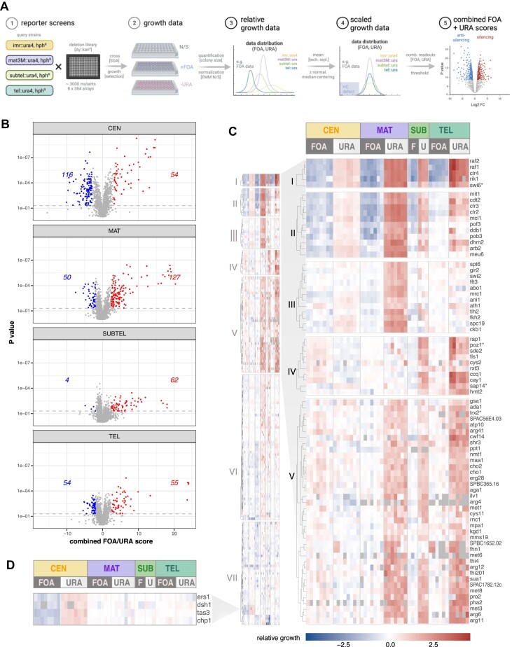
Heterochromatin plays a critical role in regulating gene expression and maintaining genome integrity. While structural and enzymatic components have been linked to heterochromatin establishment, a comprehensive view of the underlying pathways at diverse heterochromatin domains remains elusive. Here, we developed a systematic approach to identify factors involved in heterochromatin silencing at pericentromeres, subtelomeres and the silent mating type locus in Schizosaccharomyces pombe. Using quantitative measures, iterative genetic screening and domain-specific heterochromatin reporters, we identified 369 mutants with different degrees of reduced or enhanced silencing. As expected, mutations in the core heterochromatin machinery globally decreased silencing. However, most other mutants exhibited distinct qualitative and quantitative profiles that indicate heterochromatin domain-specific functions, as seen for example for metabolic pathways affecting primarily subtelomere silencing. Moreover, similar phenotypic profiles revealed shared functions for subunits within complexes. We further discovered that the uncharacterized protein Dhm2 plays a crucial role in heterochromatin maintenance, affecting the inheritance of H3K9 methylation and the clonal propagation of the repressed state. Additionally, Dhm2 loss resulted in delayed S-phase progression and replication stress. Collectively, our systematic approach unveiled a landscape of domain-specific heterochromatin regulators controlling distinct states and identified Dhm2 as a previously unknown factor linked to heterochromatin inheritance and replication fidelity.


Figures

Graphical Abstract
Graphical Abstract
Figure 1.
Figure 1.
Systematic identification of heterochromatin regulators. (A) Schematic overview of the screening strategy employing high-throughput silencing assays. The ura4+ gene in the reporter strains was positioned at the pericentromeric region (imr1L; CEN), the silent mating type locus (mat3M; MAT), the subtelomeric region (subtel2R; SUBTEL) or adjacent to telomeric repeats (tel2L; TEL). An additional hygromycin selection (hphR) marker was introduced adjacent to the heterochromatic domains. Created in BioRender. Braun, S. (2023) BioRender.com/y99c398. (B) Volcano plots illustrating the combined FOA/URA scores (x-axis) and P-values (y-axis) derived from one-sample Student’s t-test for each mutant (details in ‘Materials and methods’ section). The data include 3–8 independent biological replicates (CEN: 8; MAT: 7; SUBTEL: 3; TEL: 6), each comprising 2–4 technical replicates. Mutants with significantly altered silencing (P< 0.05) are highlighted and the respective numbers of silencing factors (right side) and anti-silencing factors factors (left side) are indicated. (C) Heatmaps displaying k-means cluster analysis of log2-transformed relative growth values (+FOA, −URA) from 176 silencing mutants exhibiting a significant decrease in heterochromatin silencing in at least one domain (details in the text). The left panel displays an overview of all clusters, while the right panel shows a subset of clusters (I–V) with gene names. The gene order within each cluster was determined through subsequent hierarchical clustering (see ‘Materials and methods’ section). (D) Heatmap showing relative growth values of a subset of genes involved in RNAi from Cluster VII. In cases where mutants were mis-annotated in the gene deletion collection, the correct gene name is indicated by an asterisk (see also Supplementary Table S8 and ‘Materials and methods’ section).
Figure 2.
Figure 2.
Similarities between phenotypic profiles reflect the composition of protein complexes involved in chromatin organization. (A) Density plot showing frequency density of Pearson correlation coefficients for the pairwise correlation of mutant profiles of 70 genes belonging to 12 complexes involved in chromatin organization (for details, see Supplementary Table S11 and the text). Lines represent gene pairs within the same or different complexes (legend is shown below graph). (B) Detailed view of pairwise correlations restricted to genes within identical complexes. Pearson correlation coefficients in (A) and (B) were calculated using average values of the combined FOA/URA score from 3 to 8 biological replicates. (C) Heatmaps showing the median log2-transformed values of the combined FOA/URA scores from 3 to 8 biological replicates (CEN: 8; MAT: 7; SUBTEL: 3; TEL: 6; each with two technical replicates) focusing on three complexes: Set1C/COMPASS (top), Mst2CNuA3 (middle) and SAGA complexes (bottom). Accompanying diagrams (left panels) depict the complex structures based on existing crystallographic data or functional genetics studies. (D) Correlation matrices highlighting Pearson correlation coefficients based on combined FOA/URA scores among subunits within each complex. (E) Expression analysis of endogenous transcripts at two heterochromatic loci (imr1L and tlh1) analyzed by RT-qPCR in selected mutants. Transcript levels are normalized against act1 levels and presented as box plots using log2-transformed values relative to the wild-type (WT) median across biological replicates (n = 4–5). Colors correspond to the different protein complex modules.
Figure 3.
Figure 3.
Metabolic pathway genes contribute to silencing at subtelomeres and mating type locus. (A) Heatmap depicting relative growth values (+FOA, −URA) of mutants specifically impaired in silencing at MAT, SUBTEL and TEL. Gene order reflects functional groups identified by GO terms. (B) Schematic representations of sulfate assimilation (top) and phosphatidylcholine synthesis (bottom) metabolic pathways. Bold protein names correspond to mutants identified under stringent selection criteria. Created in BioRender. Braun, S. (2023) BioRender.com/x78c746. (C) Quantification of heterochromatin transcript levels at cen-dg (pericentromeres), mat3M::ura4 (mating type locus) and tlh1 (subtelomeres) by RT-qPCR. Transcript levels from 3 to 4 biological replicates, normalized against act1 levels, are presented relative to the wild-type (WT) median (n = 8). Individual replicates are illustrated in a floating bar plot with the median indicated by a line. (D) Density plots displaying FC analysis of fluorescent mKO2 reporter expression at the single-cell level from a subtelomeric locus (HSSSubtel reporter system). FC experiments were conducted in mutants impaired in methionine and cysteine synthesis (top) and membrane lipid synthesis (controls: WT, dashed line; clr4Δ, solid line). The x-axis shows mKO2 reporter expression values (‘orange’) normalized against E2C expressed from a proximal euchromatic locus (noise filter, ‘red’). The y-axis represents the density of the cell population relative to the mean expression value in clr4Δ (ON state). The bottom right scheme illustrates the HSSSubtel reporter system with ‘orange’ and ‘red’ inserted at ∼28 and ∼46 kb, respectively, downstream of telomeric repeats on chromosome IIR.
Figure 4.
Figure 4.
Dhm2 contributes to heterochromatin structure and silencing at constitutive and facultative heterochromatin. (A) Multiple sequence alignment of the Dhm2 protein sequence and its homologs present in Schizosaccharomyces osmophilus, Schizosaccharomyces cryophilus and Schizosaccharomyces japonicus using the T-coffee alignment tool. Conservation levels are highlighted (dark shading indicates high conservation). (B) Predicted Dhm2 structure based on the AlphaFold2 model. (C) Silencing assay using the mat3M::ura4+ reporter. Ten-fold dilutions of wild-type (WT) cells, dhm2Δ and clr4Δ strains were plated on non-selective (N/S) and selective (lacking uracil) media. (D) Quantification of heterochromatin transcript levels at cen-dg (pericentromeric repeats), mat3M::ura4+ (mating type locus) and tlh1+ (subtelomeric gene) by RT-qPCR. Transcript levels, normalized against act1, are presented relative to the WT median value (n = 8 independent biological replicates). (E) ChIP-qPCR analysis of H3K9me2 levels at constitutive heterochromatin domains (cen-dg, mat3M::ura4+ and tlh1+) and at facultative heterochromatin islands (ssm4+, mei4+ and SPBC24C6.09c). Input-normalized IP samples are normalized to the average of two euchromatic loci (act1+ and tef3+) and shown relative to the WT median value (n = 7 and three independent biological replicates, respectively). (F) Schematic representation of heterochromatin pathways involving the RNAi machinery, SHREC and Cul4-Ddb1Cdt2. Created in BioRender. Braun, S. (2024) BioRender.com/z57f883. (G) Quantification of heterochromatic transcripts from cen-dg repeats and the mat3::ura4+ reporter gene by RT-qPCR. Transcript levels, normalized against act1, are presented relative to the WT median values (n = 3–4 independent biological replicates). (H) ChIP-qPCR analysis of H3K9me2 enrichment at cen-dg repeats and the mat3::ura4+ reporter gene. Input-normalized IP samples were normalized to the average of two euchromatic loci (act1+ and tef3+) and are shown relative to the WT median value (n = 3–4 independent biological replicates). For (D), (E), (G) and (H), the individual replicates are displayed in box whisker or floating bar plots; the line depicts the median. For (G) and (H), statistical analysis was performed using one-way ANOVA tests, with letters denoting groups with significant differences as determined by Tukey’s post-hoc tests at P < 0.05.
Figure 5.
Figure 5.
Dhm2 is required for heterochromatin maintenance. (A) Monitoring of RNAi-dependent heterochromatin establishment. Top: Schematic of the Rik1 tethering system to nascent transcripts via boxB binding sites at the 3′UTR of ura4+. Bottom: Silencing assay using the ura4+-5BoxB reporter. Serial 10-fold dilutions of strains expressing Rik1-ΔN, including wild-type (WT), positive controls (dcr1Δ, mkt1Δ) and two independent dhm2Δ strains, were plated on non-selective (N/S) and 5-FOA-containing media. The rik1+ strain expresses the non-fusion variant (negative control). (B) Monitoring of heterochromatin spreading at the silent mating type locus. Top: Schematic of the ΔREIIIHSS (heterochromatin spreading sensor) system, with reporters inserted at cenH (‘green’; nucleation site), downstream of the REIII element (‘orange’; sensor site) and mutations in the Atf1/Pcr1 binding sites of REIII element (ΔREIIIHSS) denoted by two vertical lines. An additional reporter gene (‘red’) is placed downstream of IR-R as a transcriptional noise filter (not shown). Bottom left: 2D hexbin plots display expression of ‘green’ and ‘orange’ reporters (normalized against ‘red’ expression), in WT and dhm2Δ mutant in the ΔREIIIHSS the reporter strain. Right: Density plot showing red-normalized ‘orange’ reporter expression cells filtered for ‘green’ off state. (C) Monitoring of heterochromatin establishment and maintenance at an ectopic heterochromatin locus. Top: Schematic of the inducible TetR-Clr4* establishment system at the ade6+ locus. Middle: Representative images of colony color assay. WT, dhm2Δ, epe1Δ and dhm2Δ epe1Δ carrying 4xtetO-ade6+ and expressing TetR-clr4* were grown on PMG with low adenine in the presence or absence of anhydrotetracycline (AHT). Bottom: Percentage of red-pink colonies, sectored colonies and white colonies. Mean values and the range (error bars) from two independent experiments are shown (numbers of cells examined from two experiments for -AHT: WT = 287/49, dhm2Δ = 463/480, epe1Δ = 213/108, dhm2Δ epe1Δ = 1175/605; for + ATH: WT = 511/72, dhm2Δ = 596/516, epe1Δ = 245/118, dhm2Δ epe1Δ = 458/553). (D) ChIP-qPCR analysis of H3K9me3 enrichment at ade6 locus. Input-normalized IP samples, normalized to the average of two euchromatic loci (act1+ and tef3+), and are shown relative to the WT median value in the absence of AHT (n = 3–4 independent biological replicates). Schemes in (A), (B) and (C) created in BioRender. Braun, S. (2023) BioRender.com/x35u872; Braun, S. (2024) BioRender.com/u10z983 and Braun, S. (2023) BioRender.com/h31c876, respectively.
Figure 6.
Figure 6.
Loss of Dhm2 results in induction of replication stress markers. (A) Quantification of DNA synthesis outside S-phase. DNA synthesis in non-septated cells was detected by fluorescence microscopy following a short pulse of EdU. (B) Accumulation of RPA foci. Left panel: Representative images Ssb3-YFP foci in WT and dhm2Δ cells. Right panel: Percentage of cells with RPA foci formation. Shown are mean values and errors (95% confidence interval) from 3 (WT) and 7 (dhm2Δ) biological replicates. (C) Accumulation of Rad52-GFP foci. Left panel: Representative images of WT and dhm2Δ cells expressing Rad52-GFP. Right panel: Percentage of Rad52-GFP foci formation in WT, dhm2Δ and pof3Δ cells. (D) Sensitivity to DNA damage. Ten-fold serial dilutions of the indicated strains were plated on YES medium supplemented with different DNA-damaging agents (HU, hydroxyurea; CPT, camptothecin; MMS, methyl methanesulfonate) and incubated for 3 days at 32°C. (E) RT-qPCR analysis of heterochromatic transcripts (cen-dg, mat3M::ura4+ and tlh1+) in the indicated strains. Transcript levels, normalized against act1, are presented relative to the WT median value (n = 4 independent biological replicates). (F) ChIP-qPCR analysis of H3K9me2 levels at heterochromatin domains (cen-dg, mat3::ura4+ and tlh1+) in the indicated strains. Input-normalized IP samples, normalized to the average of two euchromatic loci (act1+ and tef3+), are shown relative to the WT median value (n = 4–6 independent biological replicates). (G) Scheme illustrating factors involved in DNA replication and epigenetic inheritance. Created in BioRender. Braun, S. (2023) BioRender.com/z39c488. (H) Ascus formation efficiency and spore viability of dhm2 single and double mutants. Representative images of asci formed 3 to 4 days after mating (h+ strains, top: WT, mrc1Δ, mcl1Δ; h strains, left: WT, dhm2Δ). Table shows the genetic cross scheme (left), percentage of cells with complete tetrads (middle) and the number of tetrads with four viable spores after germination on YES media (right). For quantification of ascus formation, mean value and deviation (range) are presented from two independent experiments (∼300 cells analyzed per cross).

Update of

Similar articles

References

    1. Grewal S.I.S. The molecular basis of heterochromatin assembly and epigenetic inheritance. Mol. Cell. 2023; 83:1767–1785. - PMC - PubMed
    1. Allshire R.C., Madhani H.D.. Ten principles of heterochromatin formation and function. Nat. Rev. Mol. Cell Biol. 2018; 19:229–244. - PMC - PubMed
    1. Holoch D., Moazed D.. RNA-mediated epigenetic regulation of gene expression. Nat. Rev. Genet. 2015; 16:71–84. - PMC - PubMed
    1. Harr J.C., Gonzalez-Sandoval A., Gasser S.M.. Histones and histone modifications in perinuclear chromatin anchoring: from yeast to man. EMBO Rep. 2016; 17:139–155. - PMC - PubMed
    1. Allshire R.C., Ekwall K.. Epigenetic regulation of chromatin states in schizosaccharomyces pombe. Cold Spring Harb. Perspect. Biol. 2015; 7:a018770. - PMC - PubMed