. 2024 Dec 11;52(22):13792-13811.
doi: 10.1093/nar/gkae1136.

[SNG2], a prion form of Cut4/Apc1, confers non-Mendelian inheritance of heterochromatin silencing defect in fission yeast

Affiliations

[SNG2], a prion form of Cut4/Apc1, confers non-Mendelian inheritance of heterochromatin silencing defect in fission yeast

Suman Sharma et al. Nucleic Acids Res. .

Abstract

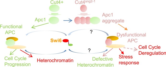
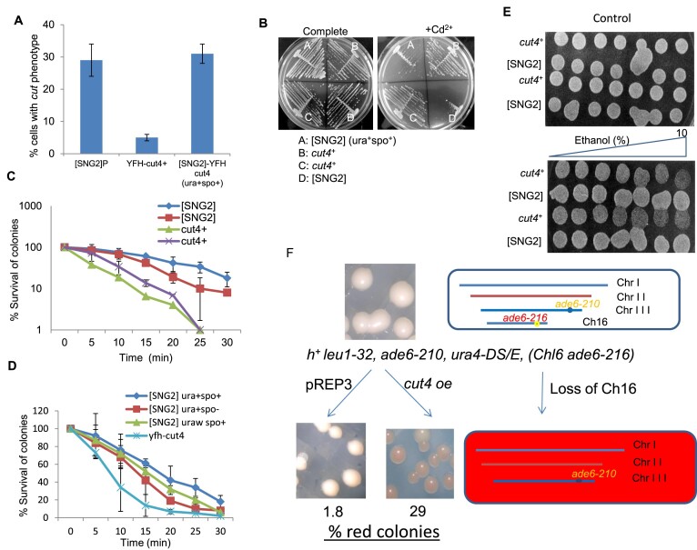
Prions represent epigenetic regulator proteins that can self-propagate their structure and confer their misfolded structure and function on normally folded proteins. Like the mammalian prion PrPSc, prions also occur in fungi. While a few prions, like Swi1, affect gene expression, none are shown to affect heterochromatin structure and function. In fission yeast and metazoans, histone methyltransferase Clr4/Suv39 causes H3-Lys9 methylation, which is bound by the chromodomain protein Swi6/HP1 to assemble heterochromatin. Earlier, we showed that sng2-1 mutation in the Cut4 subunit of anaphase-promoting complex abrogates heterochromatin structure due to defective binding and recruitment of Swi6. Here, we demonstrate that the Cut4p forms a non-canonical prion form, designated as [SNG2], which abrogates heterochromatin silencing. [SNG2] exhibits various prion-like properties, e.g. non-Mendelian inheritance, requirement of Hsp proteins for its propagation, de novo generation upon cut4 overexpression, reversible curing by guanidine, cytoplasmic inheritance and formation of infectious protein aggregates, which are dissolved upon overexpression of hsp genes. Interestingly, [SNG2] prion imparts an enhanced tolerance to stress conditions, supporting its role in promoting cell survival under environmental stress during evolution.


Conflict of interest statement

None declared.

Figures

Graphical Abstract
Graphical Abstract
Figure 1.
Figure 1.
Two alternative silencing defective states displayed by the sng2-1/cut4 mutant. (A) shows the genotype I, denoted as leu1mat1Msmto REIIΔmat2P::ura4 along with the associated phenotypes in the wt, swi6 and sng2-1 background, giving the respective iodine staining phenotypes of the colonies on minimal media (PMA+): spo, as light staining and spo+ representing dark staining. Also shown are the respective phenotypes of growth on media lacking uracil as ura or ura+, FOAr or FOAs. (B) Genotype II, denoted as mat1PΔ17::LEU2, REIIΔmat2P::ura4, with a linked his2 marker. Also shown are the phenotypes associated with WT, swi6 and sng2-1 backgrounds. (C) Spotting assay to assess the growth pattern of the control strain with genotype I along with control ura+ and ura strains on indicated plates. Also shown are the spo (LSPR) and spo+ (DSPR) derivatives of the sng2-1 mutant with genotype I. (D) RT PCR analysis to monitor the level of the polα and mat2Pc transcripts in WT (-) and LSPR and DSPR derivatives of the sng2-1 mutant. (E) Iodine phenotypes of the sng2-1 mutant in background of genotype I transformed with vector, cut4 gene in high copy vector or an integrating vector.
Figure 2.
Figure 2.
Persistence of the derepressed phenotype caused by the sng2-1 mutation after outcrossing. Tetrad analysis shows the cross between the DSPR strain with genotype I (leu1 mat1Msmto REIIΔmat2P::ura4, ura4D18, sng2-1) with a WT strain having genotype II (leu1 mat1PΔ17::LEU2, leu1-32, REIIΔmat2P::ura4, ura4D18, his2). Segregants listed as putative ‘[SNG2]M’ prion form are shown in square boxes and those as putative prion form ‘[SNG2]P are circled.
Figure 3.
Figure 3.
Non-Mendelian segregation of the putative [SNG2] prion form (A) Phenotypes of the wt, sng2-1 mutant and the putative prion derivatives, [SNG2]M and [SNG2]P, in the genetic backgrounds I and II, respectively, was monitored by spotting assay on indicated plates. (B) Tetrads derived from a cross of [SNG]P strain having spo-ura+ phenotype (top) and a WT strain (genotype I) having spo-ura phenotype (bottom), were spotted on YEA and then replica plated to different plates, as indicated. Segregants representing the putative ‘[SNG2]M’ prion form are shown in square boxes. (C) Segregation pattern of ura+ phenotypes of the mat2P::ura4 locus in tetrad analysis as shown in (B).
Figure 4.
Figure 4.
Aggregate formation by fluorescent-tagged Cut4 upon prion conversion (A) Tetrads derived from a cross between a WT strain of Minus mating type (genotype I) containing an ectopically integrated YFP-Cut4 (top) and an [SNG2]P strain with genotype II (bottom). Segregants having the putative ‘[SNG2]M’ phenotype, are shown in square boxes. (B) Confocal microscopic analysis of selected segregants from tetrads analysis shown in (A) showing YFP-Cut4 aggregation. (CF) De novo generation of prion form [SNG2]M upon overexpression of cut4. (C) Serial dilution spotting assay to show growth on plates lacking uracil and iodine staining phenotype, for transformants of WT strain (Msmto REIIΔmat2ura4) containing high copy empty vector (pREP3X), high copy vector containing full length cut4 gene or an internal deletion BglIIΔ (cut4DB/B). (D) Serial dilution spotting assay showing the persistence of elevated level of ura+ phenotype in WT strain after plasmid loss of the transformants shown in (C). (E) Quantitation of spo+ and ura+ colonies upon ectopic expression of vector, cut4 and cut4DB/B on high copy vector and upon loss of vectors, as shown in panels (A) and (B), respectively. Data was represented as standard deviation (SD). (F) Confocal microscopy pictures of cells of WT strain harboring integrated GFP tagged copy of cut4 gene upon transformation with different vectors. Numbers denote the percentage of cells containing GFP-Cut4 aggregates.
Figure 5.
Figure 5.
Curing of [SNG2] prion by Gu-HCl and overexpression of heat shock proteins. (A) Serial dilution spotting assay of DSPR, Msmto REIIΔ mat2P::ura4, SNG2]M-YFH cut4 (spo+-ura+) segregants and [SNG2]o (cut4 o/e) along with the parental strains on complete plates and plates lacking uracil, with and without treatment of 4 mM Gu-HCl. o/e denotes overexpression. (B) Quantitation of percentage of colonie with ura+ phenotype on treatment of various strains with 4 mM Gu-HCl. ura+ represents control WT, DSPR, SPA236 with genotype Msmto ura4D18 REIIΔ mat2P::ura4 and two independent [SNG2]M (spo+-ura+) segregants from Figure 2 and two independent [SNG2]0-GFP-cut4 from transformants with ura+ phenotype after plasmid loss as shown in Figure 4D. (C) Quantitation of percentage of spo+ colonies on treatment of different indicated strains with 4 mM Gu-HCl (Iodine staining indicates staining of colonies with iodine after 3 days of growth). (B, C) Data was represented as SD. (D) Curing of prion form of [SNG2]M by overexpression of hsp genes in segregant 3B (spo+-ura+) obtained from cross shown in Figure 2 by serial dilution spotting assay. (E) Quantitation of the effect of overexpression of hsp70 and hsp104 on percentage of colonies showing spo+ and ura+ phenotype. Error bars represent SD. (F) Iodine staining of transformants generated from expression of pREP3X, cut4 DB/B and cut4 in WT strain with genotype I (Msmto REIIΔmat2P::ura4) and same strain having hsp104Δ mutation (Msmto REIIΔmat2P::ura4 hsp104Δ). The transformants were streaked on selective plates (PMA-leu) and colonies stained after 4 days’ growth at 30°C.
Figure 6.
Figure 6.
Formation of Cut4 amyloid aggregates in [SNG2] prion form. (A) SDD-AGE followed by western blotting with anti-GFP antibody of the protein extracts from segregants shown in Figure 4B. Parent 1: sng2+/YFH-Cut4 (lane 1), segregant 3B, with spo+-ura+ phenotype (lane 2), segregant 2A with phenotype spo-ura+ (lane 3), segregant 2B with spo+-ura+ phenotype (lane 4), segregant 4B with spo+-uraw phenotype (lane 5) and Parent 2: [SNG2]P with no YFP-tagged Cut4 (lane 6). (B) SDD-AGE under modified conditions showing resolution of the monomeric form of Cut4 from multimeric forms. (C) Generation of aggregates of Cut4-GFP upon expression of intact but not truncated Cut4. (D) Comparison of the Cut4 prion form in the dark (D) and light (L) derivatives of the cut4sng2-1 mutant. (E) Conversion of multimeric forms of Cut4 prion form by growth in presence of guanidine. (F) Conversion of amyloid aggregates to monomeric form by overexpression of hsp104. (CE) SDD-AGE analysis of Dark and Light derivatives of sng2-1 mutant having GFP-tagged Cut4sng2-1 (C), [SNG2] cells grown in the absence and presence of guanidine (D) or hsp104 (E). (AF) Lower panels represent western blots of samples probed with anti-a-tubulin antibody. Arrowheads represent oligomeric/multimeric bands of Cut4 prion form. (G) Fractionation of sng2+ and Light (L) and Dark (D) derivatives of [SNG2] having GFP tagged Cut4. Total (T), soluble (S) and pellet (P) fractions were transferred to nitrocellulose membrane by slot blot and blot was probed with anti-GFP antibody.
Figure 7.
Figure 7.
Cytoplasmic inheritance and dominance of [SNG2]. (A) Diagrammatic representation of cytoduction experiment for generation of dominant (prion+ form) and recessive (prion form) in karyogamy deficient (tht1Δ) diploid strain. (B) Serial dilution spotting assay of strains with indicated genotypes on complete, YE, PMA+, -ura and FOA plates. Colonies growing on PMA+ plates were stained with iodine to check sporulation. Lower panel includes the control strains. Pictures on the right side and photomicrographs of the cells subjected to sporulation conditions to induce meiosis. (C) Quantitation of ura+ and spo+ colonies of strains with indicated genotypes after growth on plates lacking uracil and sporulating plates, respectively. (D) Tetrads derived from a cross of [SNG2]P-tht1Δ strain (prion form with genotype II having tht1Δ) having spo-ura+ phenotype and a WT strain with tht1Δ (genotype I) having spo-ura phenotype, were spotted on YEA and then replica plated to different plates, as indicated. (E) Segregation pattern of ura+ phenotype among tht1Δ mutants in tetrad analysis represented in (D).
Figure 8.
Figure 8.
Propagation of [SNG2] by protein transformation. (A) Diagrammatic representation of test for infectivity of [SNG2] prion, as described in the ‘Materials and methods' section and the ‘Results’ section. (B) Serial dilution spotting assay of wild-type strain (genotype I with GFP tagged cut4) transformed with protein from indicated strains, on complete PMA+, -ura and FOA plates. Colonies growing on PMA+ plates were stained with iodine to check sporulation.
Figure 9.
Figure 9.
Cells of [SNG2] prion form phenocopy cut4 mutant and show enhanced stress response. (A) Histogram representing the percentage of cells having ‘cut’ phenotype in indicated strains. (B) Growth of WT and [SNG2] segregants of a single tetrad on PMA plate containing 20 μM CdCl2. (C) Plot representing survival of WT and [SNG2] cells subjected to heat stress at 52°C for the indicated time periods. (D) Plot representing survival of WT and independent [SNG2] segregants upon exposure to 1 mM H2O2 for the indicated time periods. (E) Spotting assay of cells of two WT and [SNG2] strains each on control plate (top panel) and plate containing 0–10% gradient of ethanol (lower panel). (F) Enhanced rate of chromosome loss in [SNG2] cells. The schematic depicts the basis of screen for chromosome loss. Colonies of mutant showing enhanced loss of the artificial chromosome (Ch16 ade6-216) appeared as red colonies, while normal WT cells with high level of chromosome stability produced white colonies on the adenine-limiting YE plate (74).

Similar articles

Cited by

References

    1. Prusiner S.B. Novel proteinaceous infectious particles cause scrapie. Science. 1982; 216:136–144. - PubMed
    1. Oesch B., Westaway D., Wälchli M., McKinley M.P., Kent S.B., Aebersold R., Barry R.A., Tempst P., Teplow D.B., Hood L.E.. A cellular gene encodes scrapie PrP 27-30 protein. Cell. 1985; 40:735–746. - PubMed
    1. Moore R.C., Hope J., McBride P.A., McConnell I., Selfridge J., Melton D.W., Manson J.C.. Mice with gene targeted prion protein alterations show that Prnp, Sinc and Prni are congruent. Nat. Genet. 1998; 18:118–125. - PubMed
    1. Prusiner S.B. Prions. Proc. Natl Acad. Sci. U.S.A. 1998; 95:13363–13383. - PMC - PubMed
    1. Zabel M.D., Reid C.. A brief history of prions. FEMS Path Disease. 2015; 73:ftv087. - PMC - PubMed

MeSH terms

LinkOut - more resources