. 2024 Dec 11;52(22):13577-13593.
doi: 10.1093/nar/gkae1097.

Glucose binds and activates NSUN2 to promote translation and epidermal differentiation

Affiliations

Glucose binds and activates NSUN2 to promote translation and epidermal differentiation

Weili Miao et al. Nucleic Acids Res. .

Abstract

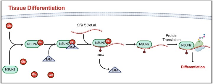
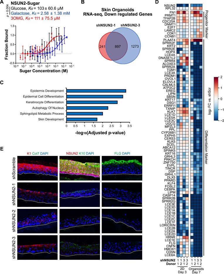
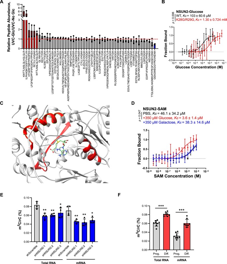
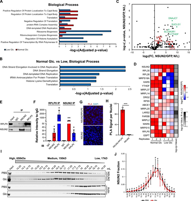
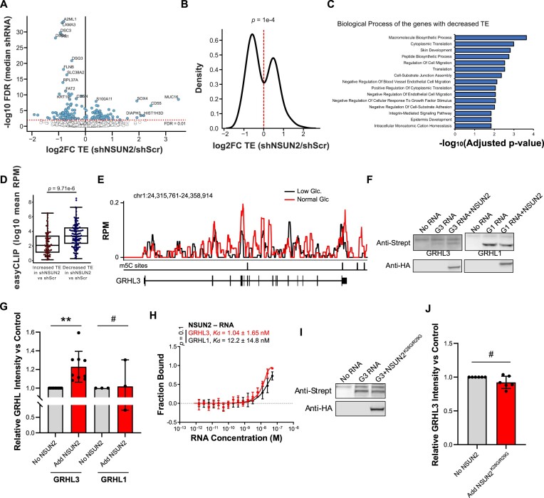
Elevations in intracellular glucose concentrations are essential for epithelial cell differentiation by mechanisms that are not fully understood. Glucose has recently been found to directly bind several proteins to alter their functions to enhance differentiation. Among the newly identified glucose-binding proteins is NSUN2, an RNA-binding protein that we identified as indispensable for epidermal differentiation. Glucose was found to bind conserved sequences within NSUN2, enhancing its binding to S-adenosyl-L-methionine and boosting its enzymatic activity. Additionally, glucose enhanced NSUN2's proximity to proteins involved in mRNA translation, with NSUN2 modulating global messenger RNA (mRNA) translation, particularly that of key pro-differentiation mRNAs containing m5C modifications, such as GRHL3. Glucose thus engages diverse molecular mechanisms beyond its energetic roles to facilitate cellular differentiation processes.


Figures

Graphical Abstract
Graphical Abstract
Figure 1.
Figure 1.
Glucose binds to NSUN2 and NSUN2 is essential for differentiation. (A) MST quantifies NSUN2’s binding affinity for glucose, 3OMG and galactose. (B) Overlap of downregulated genes (FDR < 0.05) between the two shRNAs for NSUN2 versus shScramble in RNA-seq of human epidermal organoid tissues. (C) Biological process GO terms for RNAs downregulated upon NSUN2 depletion (the 897 gene intersection in Figure 1B). (D) Changes in progenitor and differentiation markers upon NSUN2 loss in 2D day 3 differentiated keratinocytes and in skin organoid tissues (RNA-seq); differentiation markers shown include all genes with a GO term of keratinocyte differentiation, regulation of keratinocyte differentiation, or keratinization, with a fold-change >4 in any biological replicate. (E) Keratin 1 (K1), Keratin 10 (K10), Filaggrin (FLG), NSUN2 and Col7 immunostaining in regenerated human skin organoid tissues, treated with NSUN2 shRNAs or control shRNA; dotted line, basement membrane. Error bars = S.D. from ≥3 biological replicates.
Figure 2.
Figure 2.
Glucose binding to NSUN2 requires amino acids K28/R29. (A) Relative abundance of the peptides with no glucose modification, calculated from the intensity obtained from 1 mM glucose/no-glucose, both with UVC crosslinking. The peptides with increased, no changes and decreased abundance after glucose treatment has been labeled with different colors. (B) MST of NSUN2 WT and NSUN2K28G/R29G mutant binding affinity for glucose. (C) Molecular docking by UCSF Chimera shows the predicted binding site of SAM to NSUN2 (structure: AF-Q08J23-F1 from AlphaFold). Peptide sequences with increased accessibility were highlighted in the structure. (D) MST of NSUN2 binding affinity for SAM ± 350 μM glucose or galactose. (E) Percentage of m5C/rC with NSUN2 shRNAs and control shRNA treatment from two biological replicates and two technical replicates. (F) Percentage of m5C/rC in keratinocyte progenitor cells (Prog.) and day 3 differentiated cells (Diff.) from four biological replicates and two technical replicates. Error bars = S.D. from ≥3 biological replicates if not notified specifically. *, 0.01 < P≤ 0.05; **, 0.001 < P≤ 0.01; ***, P≤ 0.001.
Figure 3.
Figure 3.
Glucose regulates NSUN2 binding to specific pro-differentiation mRNA. (A) Motif identified by HOMER de novo searching of NSUN2 CLIP-seq peaks. (B) Binding of NSUN2 to mRNA exons or introns in undifferentiated (Prog.) and differentiated human keratinocytes grown in low (Low Glc.) or normal glucose (Normal Glc.). (C) Heatmap of the binding of NSUN2 to the NSUN2-mediated m5C levels of known RNAs in differentiated keratinocytes grown in normal versus low glucose (N/L). (D) Binding traces of NSUN2 to RNY1 RNA in differentiated keratinocytes grown in low or normal glucose from two independent donors. (E) Heatmap of the binding of NSUN2 to pro-differentiation mRNAs in the exons or introns in differentiated keratinocytes grown in normal versus low glucose (N/L). (F) Binding traces of NSUN2 to JUP, SPRR1B and S100A9 mRNAs in differentiated keratinocytes grown in low or normal glucose. Error bars, standard deviations from ≥3 biological replicates. **, 0.001 < P≤ 0.01; ***, P≤ 0.001.
Figure 4.
Figure 4.
Glucose promotes NSUN2 binding to translation-related complex. (A) Adjusted P-values of top biological process GO terms for the genes enriched in NSUN2 proximal proteomes in low or normal glucose conditions compared to corresponding GFP. (B) Adjusted P-values of top biological process GO terms for the genes enriched in NSUN2 proximal proteomes in normal glucose conditions compared to low glucose using the ratios of NSUN2 bioID normalized to the corresponding GFP bioID. (C) Volcano plot of the protein enrichment in normal glucose versus low glucose of NSUN2 bioID (normalized to corresponding GFP bioID) of the 254 enriched proteins. Some of the translation-related proteins and some other noteworthy proteins were highlighted. (D) Heatmap of the translation-related proteins enriched in NSUN2 proximal proteomes in low glucose or normal glucose conditions. (E) Western blot of NSUN2 and RPLP0 co-IP in day 3 differentiated cells. (F) Relative co-purification enrichment in NSUN2 and RPLP0 co-IP normalized to IgG control. (G) PLA signal for the interaction between NSUN2 and RPLP0 in differentiated cells maintained in normal glucose. (H) Quantitative analysis of PLA signal. (I) Western blot of NSUN2 and β-actin in FPLC from 14.25 to 19.25 ml, ±1 mM glucose incubation in the cell lysates. (J) Distribution of NSUN2 protein in FPLC, ±1 mM glucose in the cell lysates, from western blot. Error bars, standard deviations from ≥3 biological replicates. #, P> 0.05; *, 0.01 < P≤ 0.05; **, 0.001 < P≤ 0.01; ***, P≤ 0.001.
Figure 5.
Figure 5.
Glucose regulates NSUN2 and drives translation of pro-differentiation mRNAs. (A) Volcano plot of TE differences between shNSUN2 and shScramble. (B) Kernel density smoothed histogram of the change in translational efficiency upon NSUN2 knock-down for mRNA with significant efficiency changes (FDR < 0.1), but limited change in mRNA levels (|log2|<0.5). mRNAs with large changes in abundance upon NSUN2 loss were removed to avoid possible technical biases. P-value represents the change in TE in significant genes being different from all genes. (C) Adjusted P-values of top biological process GO terms for the genes with significantly decreased TE (FDR < 0.1) in shNSUN2 versus shScramble and limited (|log2|<0.5) mRNA level changes. (D) Box and scatter plots of the mean reads from easyCLIP-seq in day 3 differentiated cells maintained in normal glucose for RNAs with have significantly decreased TE (FDR < 0.1) in shNSUN2 versus shScramble and limited (|log2|<0.5) mRNA level changes (P-value, two-sided Mann–Whitney). (E) Binding traces of NSUN2 across GRHL3 mRNA in differentiated keratinocytes grown in low (black) or normal (red) glucose. (F) Western blot analysis of anti-Streptavidin representing GRHL1 or GRHL3 proteins, with or without adding FHH-NSUN2, from in vitro translation. (G) Quantitative analysis of the western blot result of GRHL1 (n = 3) and GRHL3 (n = 8) intensities from in vitro translation, normalizing to the condition without FHH-NSUN2 protein. (H) MST of NSUN2 binding to GRHL1 or GRHL3 full-length mRNAs. (I) Western blot analysis of anti-Streptavidin, representing GRHL3 protein with or without adding FHH-NSUN2K28G/R29G. (J) Quantitative analysis of the western blot of GRHL3 intensity, normalizing to the condition without FHH-NSUN2K28G/R29G protein (n = 5). Error bars, standard deviations from ≥3 biological replicates. #, P> 0.05; **, 0.001 < P≤ 0.01.

References

    1. Blanpain C., Horsley V., Fuchs E.. Epithelial stem cells: turning over new leaves. Cell. 2007; 128:445–458. - PMC - PubMed
    1. Alonso L., Fuchs E.. Stem cells of the skin epithelium. Proc. Natl Acad. Sci. USA. 2003; 100:11830–11835. - PMC - PubMed
    1. Lopez-Pajares V., Yan K., Zarnegar B.J., Jameson K.L., Khavari P.A.. Genetic pathways in disorders of epidermal differentiation. Trends Genet. 2013; 29:31–40. - PMC - PubMed
    1. Bikle D.D., Xie Z., Tu C.-L.. Calcium regulation of keratinocyte differentiation. Exp. Rev. Endocrinol. Metabol. 2012; 7:461–472. - PMC - PubMed
    1. Fuchs E. Scratching the surface of skin development. Nature. 2007; 445:834–842. - PMC - PubMed

MeSH terms

LinkOut - more resources