. 2024 Nov 4;65(13):43.
doi: 10.1167/iovs.65.13.43.

The Mechanisms of Neuroprotection by Topical Rho Kinase Inhibition in Experimental Mouse Glaucoma and Optic Neuropathy

Affiliations

The Mechanisms of Neuroprotection by Topical Rho Kinase Inhibition in Experimental Mouse Glaucoma and Optic Neuropathy

Sarah E Quillen et al. Invest Ophthalmol Vis Sci. .

Abstract

Purpose: The purpose of this study was to delineate the neuroprotective mechanisms of topical 2% ripasudil (Rip), a Rho kinase (ROCK) inhibitor.

Methods: In 340 mice, scheduled 2% Rip or balanced salt solution (BSS) saline drops were intermittently, unilaterally delivered. Intracameral microbead glaucoma (GL) injection increased intraocular pressure (IOP) from 1 day to 6 weeks (6W), whereas other mice underwent optic nerve (ON) crush. Retinal ganglion cell (RGC) loss was assessed using retinal wholemount anti-RNA Binding Protein with Multiple Splicing (RBPMS) labeling and ON axon counts. Axonal transport was quantified with β-amyloid precursor protein (APP) immunolocalization. Micro-Western (Wes) analysis quantified protein expression. Immunofluorescent expression of ROCK pathway molecules, quantitative astrocyte structural changes, and ON biomechanical strains (explanted eyes) were evaluated. ROCK activity assays were conducted in separate ON regions.

Results: At 6W GL, mean RGC axon loss was 6.6 ± 13.3% in Rip and 36.3 ± 30.9% in BSS (P = 0.04, n = 10/group). RGC soma loss after crush was lower with Rip (68.6 ± 8.2%) than BSS (80.5 ± 5.7%, P = 0.006, n = 10/group). After 6W GL, RGC soma loss was lower with Rip (34 ± 5.0%) than BSS (51 ± 8.1%, P = 0.03, n = 10/group). Axonal transport of APP within the unmyelinated ON (UON) was unaffected by Rip. Maximum principal mechanical strains increased similarly in Rip and BSS-treated mice. Retinal ROCK 1 and 2 activity was reduced by Rip in GL eyes. The pROCK2/ROCK2 protein ratio rose in the retina of BSS GL eyes, but not in Rip GL eyes.

Conclusions: Topical Rip reduced RGC loss in GL and ON crush, with suppression of ROCK signaling in the retina and ON. The neuroprotection mechanisms appear to involve effects on both RGC and astrocyte responses to IOP elevation.


Conflict of interest statement

Disclosure: S.E. Quillen, None; E.C. Kimball, None; K.A. Ritter-Gordy, None; L. Du, None; Z. Yuan, None; M.E. Pease, None; S. Madhoun, None; T.D. Nguyen, None; T.V. Johnson, None; H.A. Quigley, None; I.F. Pitha, None

Figures

Figure 1.
Figure 1.
APP analysis using median brightness and mean >97.5 percentile pixel brightness. (A) Median APP brightness and (B) mean >97.5 percentile by region for each treatment group. APP median brightness was highest in retina and declined sequentially from prelamina to UON to MON. Median brightness in prelamina and UON was maintained at a higher level in the fellow eyes of ripasudil-treated GL mice than in fellow eyes of BSS-treated GL mice. The mean brightness values >97.5 percentile were higher in GL eyes than fellow eyes in both ripasudil and BSS treated eyes in UON, indicating axonal transport blockade in both groups. Naïve = bilaterally untreated naïve, RF = ripasudil fellow eye, R GL = ripasudil GL eye, BSS F = BSS fellow eye, BSS G = BSS GL eye. P values, ANOVA, * P ≤ 0.05, *** P ≤ 0.001.
Figure 2.
Figure 2.
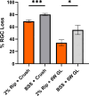
Percent RGC cell density loss by RPBMS labeling for 2W ON crush and 6W GL groups. Percent RCG soma loss with eyes treated with either ripasudil or saline drops with 6W GL or 2W crush. Group means with standard errors. * P ≤ 0.05, *** P ≤ 0.006, Wilcoxon rank sum test compared to contralateral eyes.
Figure 3.
Figure 3.
ROCK activity 24 hours after GL. Legend: Ratio comparisons with naïve eyes to two GL groups and ripasudil alone treated eyes. N = 10, t-test, * P ≤ 0.05, ** P ≤ 0.01 for comparison of each group to bilaterally untreated control eyes (equal to 1).
Figure 4.
Figure 4.
Strain responses of astrocytic lamina in eGFP mice comparing 4 groups; naïve, ripasudil alone, ripasudil GL, and BSS GL. Strain analysis (Emax and γmax) for various treatment groups and control samples; bilaterally untreated naïve, ripasudil only, and GL samples BSS + 3D GL, Ripasudil + 3D GL. Significant difference between bilaterally naïve both GL groups for both strains.
Figure 5.
Figure 5.
Labeling of ROCK pathway molecules in controls, BSS GL, and ripasudil GL eyes. Protein immunofluorescence for various treatment groups and control samples; bilaterally untreated naïve (AE) and GL samples: BSS + 3D GL (FJ), Rip + 3D GL (KO). Scale bar = 100 um.
Figure 6.
Figure 6.
Protein quantification by Wes analysis. Protein analysis for various treatment and control groups; bilaterally untreated naïve, ripasudil (cross hatched bars) only, and 3-day pressure elevated samples treated with either ripasudil (orange) or BSS (grey) drops. Comparison of the ratios of ROCK species (AC): proteins RhoA, ROCK1, ROCK2, and pROCK2, as well as downstream proteins (DF): cofilin, p-cofilin, and p38 MAPK, normalized to GAPDH level in the retina (A, D), UON (B, E) and MON (C, F), and presented as a ratio to the bilaterally untreated samples. All samples were compared to the bilaterally naïve samples with a ratio of 1. * P ≤ 0.05, † P ≤ 0.01, ‡ P ≤ 0.001, § P ≤ 0.0001, t-test.
Figure 7.
Figure 7.
Semiquantitative grading of p-ROCK1 images from UON in ripasudil GL and BSS GL. Semiquantitative grading of the intensity of the label for pROCK1.

References

    1. Rao VP, Epstein DL.. Rho GTPase/Rho kinase inhibition as a novel target for the treatment of glaucoma. BioDrugs. 2007; 21(3): 167–177. - PubMed
    1. Gonzalez LE, Boylan PM.. Netarsudil for the treatment of open-angle glaucoma and ocular hypertension: a literature review. Ann Pharmacother. 2021; 55(8): 1025–1036. - PubMed
    1. Shiuey EJ, Mehran NA, Ustaoglu M, et al. .. The effectiveness and safety profile of netarsudil 0.02% in glaucoma treatment: real-world 6-month outcomes. Graefes Arch Clin Exp Ophthalmol. 2022; 260(3): 967–974. - PubMed
    1. Yamamoto K, Maruyama K, Himori N, et al. .. The novel Rho kinase (ROCK) inhibitor K-115: a new candidate drug for neuroprotective treatment in glaucoma. Invest Ophthalmol Vis Sci. 2014; 55(11): 7126–7136, PMID: 25277230. - PubMed
    1. Nishijima E, Namekata K, Kimura A, et al. .. Topical ripasudil stimulates neuroprotection and axon regeneration in adult mice following optic nerve injury. Sci Reports. 2020; 10: 15709. - PMC - PubMed

MeSH terms