. 2024 Dec 20;13(12):3926-3935.
doi: 10.1021/acssynbio.4c00265. Epub 2024 Nov 20.

CRISPR Diagnostics for Quantification and Rapid Diagnosis of Myotonic Dystrophy Type 1 Repeat Expansion Disorders

Affiliations

CRISPR Diagnostics for Quantification and Rapid Diagnosis of Myotonic Dystrophy Type 1 Repeat Expansion Disorders

Koji Asano et al. ACS Synth Biol. .

Abstract

Repeat expansion disorders, exemplified by myotonic dystrophy type 1 (DM1), present challenges in diagnostic quantification because of the variability and complexity of repeat lengths. Traditional diagnostic methods, including PCR and Southern blotting, exhibit limitations in sensitivity and specificity, necessitating the development of innovative approaches for precise and rapid diagnosis. Here, we introduce a CRISPR-based diagnostic method, REPLICA (repeat-primed locating of inherited disease by Cas3), for the quantification and rapid diagnosis of DM1. This method, using in vitro-assembled CRISPR-Cas3, demonstrates superior sensitivity and specificity in quantifying CTG repeat expansion lengths, correlated with disease severity. We also validate the robustness and accuracy of CRISPR diagnostics in quantitatively diagnosing DM1 using patient genomes. Furthermore, we optimize a REPLICA-based assay for point-of-care-testing using lateral flow test strips, facilitating rapid screening and detection. In summary, REPLICA-based CRISPR diagnostics offer precise and rapid detection of repeat expansion disorders, promising personalized treatment strategies.

Keywords: CRISPR diagnostics; CRISPR-Cas3; Myotonic dystrophy type 1; genetic diagnosis; quantitative detection; triplet repeat diseases.


Conflict of interest statement

The authors declare no competing financial interest.

Figures
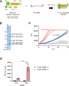
Figure 1
Figure 1
In vitro collateral cleavage activity of CRISPR-Cas3 for CTG repeats. (A) Schematic representation of the in vitro assembly of the Cascade-crRNA complex. pre-crRNA was added to a mixture of Cascade components containing Cas5, Cas6, Cas7, Cas8, and Cas11 proteins, and incubated at 37 °C for 10 min. (B) Each of Cas5, Cas6, Cas7, Cas8, and Cas11 were expressed using the baculovirus expression system in Sf9 insect cells. M: protein marker. (C) Time courses of collateral ssDNA cleavage activity measured by incubation of E. coli Cas3 and Cascade with or without a 60-bp dsDNA activator containing a target sequence containing CTG (CAG) repeats and an FQ-labeled ssDNA probe in reaction buffer containing MgCl2, CoCl2, and ATP for 30 min at 37 °C. (D) CRISPR-Cas3-mediated collateral ssDNA cleavage activity at 10 min after targeting CTG (CAG) repeat-dsDNA in fragments, quantitatively represented by relative fluorescence units (RFU). Means (n = 3), and standard deviations. ***p < 0.001, one-way ANOVA with posthoc test.
Figure 2
Figure 2
Quantitative detection of CTG repeats by CRISPR-Cas3. (A) Illustration showing the concept of the detection of CTG repeat expansion by CRISPR-Cas3. More Cascade complexes bind DNA from DM1 patients with CTG repeat expansion, resulting in a higher intensity ssDNA cleavage signal. Conversely, in unaffected individuals with less than 35 CTG repeats, only a very low cleavage signal is obtained. (B) Quantitative assay detecting plasmids containing exons 11 to 15 of the DMPK gene and CTG repeats of 0, 12 (unaffected), 240, 480, or 960 (patient-like) (left). ssDNA cleavage signals are represented by RFU per min; graph shows increasing rate of RFU/min (right). Means (n = 3), and standard deviations. *p < 0.05, **p < 0.01, one-way ANOVA with posthoc test.
Figure 3
Figure 3
Quantitative detection of CTG repeats in DM1 patients using the REPLICA method. (A) Illustration showing the concept of the REPLICA assay. Quantitative detection of repeats is achieved by detecting amplified products containing various lengths of CTG repeats from the CTG repeat region of the DMPK gene locus using REPLICA, obtained by triplet repeat-primed PCR (RP-PCR). (B) REPLICA assay from genomic DNA of DM1 patients with various CTG repeats or from genomic DNA of unaffected individuals. (C) Correlation between cleavage signal in the REPLICA assay and CTG repeat number up to 1000 repeats. (D) Cleavage signal in the REPLICA assay for Coriell samples classified by symptoms (healthy, mild, classical, or congenital) based on available clinical information. (E) REPLICA assay for detecting clinical samples from three DM1 patients or 30 non-DM1 individuals. ssDNA cleavage signals are represented by RFU per min; graph shows increasing rate of RFU/min (right). Means (n = 3), and standard deviations. *p < 0.05, ***p < 0.001, one-way ANOVA with posthoc test.
Figure 4
Figure 4
RPA-REPLICA for point of care testing (POCT) for DM1. (A) Schematic diagram of RPA-REPLICA for POCT. Amplification of various lengths of CTG repeat regions at isothermal conditions of 37 °C using RPA, followed by an ssDNA cleavage assay with REPLICA, and detection of a positive band indicating repeat expansion in approximately 5 min using a test strip. (B) Detection of repeats using a test strip. The red arrow indicates the positive band for CTG repeats. T, test band; C, control band. (C,D) Detection of CTG repeats in RPA-REPLICA for POCT. NC, negative control; HC, healthy control. (E) Detection of amplified CTG repeats with RPA-REPLICA. (F) Comparison between CTG repeat lengths and ssDNA cleavage signals.

Similar articles

Cited by

References

    1. Clark M. M.; Stark Z.; Farnaes L.; Tan T. Y.; White S. M.; Dimmock D.; Kingsmore S. F. Meta-analysis of the diagnostic and clinical utility of genome and exome sequencing and chromosomal microarray in children with suspected genetic diseases. NPJ. Genom Med. 2018, 3, 16.10.1038/s41525-018-0053-8. - DOI - PMC - PubMed
    1. Miller D. T.; Adam M. P.; Aradhya S.; Biesecker L. G.; Brothman A. R.; Carter N. P.; Church D. M.; Crolla J. A.; Eichler E. E.; Epstein C. J.; Faucett W. A.; Feuk L.; Friedman J. M.; Hamosh A.; Jackson L.; Kaminsky E. B.; Kok K.; Krantz I. D.; Kuhn R. M.; Lee C.; Ostell J. M.; Rosenberg C.; Scherer S. W.; Spinner N. B.; Stavropoulos D. J.; Tepperberg J. H.; Thorland E. C.; Vermeesch J. R.; Waggoner D. J.; Watson M. S.; Martin C. L.; Ledbetter D. H. Consensus statement: chromosomal microarray is a first-tier clinical diagnostic test for individuals with developmental disabilities or congenital anomalies. Am. J. Hum. Genet. 2010, 86 (5), 749–764. 10.1016/j.ajhg.2010.04.006. - DOI - PMC - PubMed
    1. Kamsteeg E. J.; Kress W.; Catalli C.; Hertz J. M.; Witsch-Baumgartner M.; Buckley M. F.; van Engelen B. G.; Schwartz M.; Scheffer H. Best practice guidelines and recommendations on the molecular diagnosis of myotonic dystrophy types 1 and 2. Eur. J. Hum Genet 2012, 20 (12), 1203–1208. 10.1038/ejhg.2012.108. - DOI - PMC - PubMed
    1. Radvansky J.; Ficek A.; Minarik G.; Palffy R.; Kadasi L. Effect of unexpected sequence interruptions to conventional PCR and repeat primed PCR in myotonic dystrophy type 1 testing. Diagn Mol. Pathol 2011, 20 (1), 48–51. 10.1097/PDM.0b013e3181efe290. - DOI - PubMed
    1. Shelbourne P.; Davies J.; Buxton J.; Anvret M.; Blennow E.; Bonduelle M.; Schmedding E.; Glass I.; Lindenbaum R.; Lane R.; et al. Direct diagnosis of myotonic dystrophy with a disease-specific DNA marker. N. Engl. J. Med. 1993, 328 (7), 471–475. 10.1056/NEJM199302183280704. - DOI - PubMed

Publication types

MeSH terms

LinkOut - more resources