. 2024 Nov 22;10(47):eadq4785.
doi: 10.1126/sciadv.adq4785. Epub 2024 Nov 20.

A potent proresolving mediator 17R-resolvin D2 from human macrophages, monocytes, and saliva

Affiliations

A potent proresolving mediator 17R-resolvin D2 from human macrophages, monocytes, and saliva

Mélissa Simard et al. Sci Adv. .

Abstract

Production of specialized proresolving mediators (SPMs) during the resolution phase in the acute inflammatory response is key to orchestrating complete resolution. Here, we uncovered a trihydroxy resolvin in fresh human saliva. We identified and determined its complete stereochemistry as 7S,16R,17R-trihydroxy-4Z,8E,10Z,12E,14E,19Z-docosahexaenoic acid (17R-RvD2) using total organic synthesis and matching of physical properties. The 17R-RvD2 was produced by activated human M2-like macrophages, M1-like macrophages, and human peripheral blood monocytes. 17R-RvD2 displayed potent proresolving functions (picomolar to nanomolar). Topical application of 17R-RvD2 on mouse ear skin reduced neutrophilic infiltration (~50%). 17R-RvD2 increased M2 markers CD206 and CD163 on human monocyte-derived macrophages and enhanced efferocytosis of senescent red blood cells by M2-like macrophages (EC50 ~ 2.6 × 10⁻¹⁴ M). In addition, 17R-RvD2 activated the RvD2 receptor and was equipotent to its epimer RvD2. 17R-RvD2 also significantly increased phagocytosis of Escherichia coli by human neutrophils. Together, these results establish the complete stereochemistry and potent proresolving functions of the previously unknown 17R-RvD2.


Figures
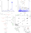
Fig. 1.
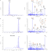
Fig. 1.. A new epimeric RvD2 product in human saliva.
(A) (Left) Representative MRM for RvD2 m/z 375 > 141 in synthetic RvD2 (black) and saliva (blue). (Right) Enhanced product ion (EPI) spectrum of the new Rv in saliva. Red arrow denotes Q3 transition daughter ion (B) Stereospecific total organic synthesis of 17R-RvD2. The synthesis of optically pure 17R-Rv D2 was accomplished by a Sonogashira coupling of the chiral vinyl iodide I with the chiral alkyne II in tert-butylamine to give the protected alkyne III. Mild deprotection of the TBS and isopropylidene groups with 1N HCl produced intermediate IV. Selective cis-reduction of the triple bond in IV with the Boland Zn/Cu/Ag alloy at 40°C gave the methyl ester of 17R-RvD2 (V). Hydrolysis of the methyl ester V with 1N LiOH afforded the target product 17R-RvD2. (C) Full two-dimensional 1H-1H correlation spectroscopy (COSY) NMR and UV spectra, together unambiguously confirming the structure of synthetic 17R-RvD2.
Fig. 2.
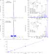
Fig. 2.. Direct comparison of physical properties of 17R-RvD2 and RvD2 and matching in human saliva.
(A) (Left) Representative MRM for synthetic RvD2 m/z 375 > 141 and (right) its MS/MS spectrum. (B) (Left) Representative MRM for synthetic 17R-RvD2 m/z 375 > 141 and (right) its MS/MS spectrum. (C) (Left) Representative MRM of 17R-RvD2 m/z 375 > 141 identified in saliva and (right) its MS/MS spectrum. Data were collected, analyzed, and presented as screen captures using Sciex OS-Q version 3.1.5.3945 (Sciex). The default values of 0.00th for m/z are depicted in figures. Note the accuracy of this instrument is approximately ± m/z 0.1.
Fig. 3.
Fig. 3.. Identification of 17R-RvD2 in human saliva using DMED derivatization.
(A) (Left) MRM chromatogram of 17R-RvD2-DMED standard with retention time (TR) = 6.15 min. (Right) Corresponding MS/MS product ion spectrum and proposed fragmentation. (B) DMED-derivatized 17R-RvD2 in human saliva with (left) chromatographic retention time at 6.14 min and corresponding (right) MS/MS spectrum run in positive mode. (C) Calibration curve of 17R-RvD2-DMED showing a linear fit r2 value of 0.999 from triplicate injections.
Fig. 4.
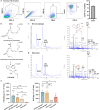
Fig. 4.. Production of 17R-RvD2 by human macrophages and monocytes.
(A) Representative flow cytometry gating strategy identifying neutrophils as the predominant leukocyte subtype in human saliva. (B) Schematic overview of 17R-RvD2 proposed biosynthesis. DHA is converted to 17R-HpDHA, which is converted by human 5-LOX into 17R-RvD2. (C to E) Monocyte-derived M1-like and M2-like macrophages, monocytes, and neutrophils were incubated with 5 μg of DHA (or vehicle) and zymosan (100 ng/ml) for 40 min. Conversion of DHA into 17R-RvD2 and RvD2 was measured using LC-MS/MS. (C) (Left) Representative MRM for 17R-RvD2 m/z 375 > 141 and (right) EPI spectra in M2-macrophage incubations. (D) (Left) Representative MRM for 17R-RvD2 m/z 375 > 141 and (right) EPI spectra in monocyte incubations. (E) Conversion of DHA into 17R-RvD2 and RvD2 by human leukocytes. Values represent the mean ± SEM, n = 3 to 4 healthy donors. Two-way ANOVA with Tukey’s multiple comparisons test, *P < 0.05, **P < 0.01, ***P < 0.001, and ****P < 0.0001; comparison of the incubation with 5 μg of DHA versus vehicle ##P < 0.01 and ####P < 0.0001.
Fig. 5.
Fig. 5.. 17R-RvD2 displays proresolving activity.
(A) Mouse ears were topically treated with 1 μg of 17R-RvD2 or vehicle (acetone, 20 μl) and inflammation was induced 7 min later by topical application of 1 μg of LTB4 plus 1 μg of PGE2. After 24 hours, the ears were digested, and leukocytes were identified by flow cytometry. (B) Representative flow cytometry gating strategy for leukocytes (LTB4 + PGE2 + vehicle). (C) Number of neutrophils (Ly6G+ cells) in the mouse ear 24 hours after the application of 1 μg of 17R-RvD2 treatments (n = 3 mice). (D) Percent of neutrophils (Ly6G+ cells) in the mouse ear 24 hours after the application of the treatments. Results are represented as mean ± SEM. n = 3 mice. Statistical analysis was carried out using Student’s t test. *P < 0.05. (E) Schematic overview of monocyte-derived M2-like macrophage generation: PBMCs were isolated from human peripheral blood and plated into six-well plates (30 × 106 PBMCs per well), which were washed to remove nonadherent cells. Remaining monocytes were differentiated into macrophages using media with MCSF (20 ng/ml) for 7 days and polarized into M2-like macrophages using media with IL-4 (20 ng/ml) for 48 hours. (F) Representative flow cytometry histogram of M2-like macrophage markers, CD163 and CD206, showing the effect of 1 nM 17R-RvD2 or corresponding amount of vehicle on the polarization of macrophages. (G) Percentage of CD206 and CD163 double-positive macrophages. Results are represented as mean ± SEM. n = 5 healthy human donors. Student’s t test, *P < 0.05 compared to IL-4 plus vehicle (H to I) Dose response: modulation by 17R-RvD2 (10−15 to 10−12 M) of CFSE-labeled senescent RBCs efferocytosis by M2 macrophages (ratio of 1 macrophage: 50 RBCs). The EC50 was estimated using nonlinear regression (dashed blue line) with log (17R-RvD2) versus response (three parameters). Results are represented as mean ± SEM. n = 5 healthy human donors. One-way ANOVA with Bonferroni multiple comparisons test, *P < 0.05 compared to M2 with RBC and vehicle.
Fig. 6.
Fig. 6.. 17R-RvD2 activates the GPR18 receptor.
(A and B) Impedance changes across CHO cell monolayers (1 × 10⁵ cells per 16-well E-plates) treated with 17R-RvD2, RvD2 (1 to 10 nM), or vehicle alone were continuously recorded each 10 s in real time for 10 min at 37°C. (A) Representative real-time recordings of impedance, comparing RvD2 vs. 17R-RvD2 at 1 nM. (B) Dose responses: RvD2- and 17R-RvD2-induced changes of impedance from 0 to 10 min are expressed as changes of cell index. Results are mean ± SEM from three independent experiments; one-way ANOVA with Tukey’s multiple comparisons test. *P < 0.05 and **P < 0.01 compared to vehicle. (C and D) CHO cells (2×10⁵ cells) were incubated with IBMX (50 μM) for 10 min, followed by addition of RvD2, 17R-RvD2, or vehicle for 15 min and cAMP levels measured by ELISA. Results are expressed as fold changes of vehicle control; mean ± SEM, n = 4 and duplicates in each experiment. (C) Dose responses: Production of cAMP in CHO-hGPR18 cells incubated with the Rvs (0.1 to 100 nM) compared to vehicle. The EC50 was estimated using nonlinear regression (dashed lines) with log (17R-RvD2) versus response (three parameters). Statistical analysis was carried out using one-way ANOVA with Dunnett’s multiple comparisons test. *P < 0.05 and **P < 0.01 compared with vehicle. (D) Comparison of cAMP produced by CHO-WT cells and CHO-hGPR18 cells treated with 1 nM RvD2 or 17R-RvD2. One-way ANOVA with Dunnett’s multiple comparisons test. *P < 0.05 compared to CHO-WT.
Fig. 7.
Fig. 7.. 17R-RvD2 increases neutrophil phagocytosis of E. coli.
(A) Representative time-course of BacLight Green–labeled E. coli phagocytosis by human neutrophils (ratio of 1 neutrophil: 50 E. coli) pre-incubated for 15 min with 1 and 10 nM 17R-RvD2. (B) Percentage increase of E. coli phagocytosis above the vehicle after 45 min in neutrophils incubated with 10 nM 17R-RvD2, RvD2, and GPR18-specific agonist or vehicle. Results are represented as mean ± SEM. n = 3 healthy human donors. Student’s t test compared to vehicle, *P < 0.05 and **P < 0.01 compared to E.coli plus vehicle. One-way ANOVA with Tukey’s multiple comparisons test was used to compare conditions. (C) Percent increase of intracellular ROS in neutrophils pre-incubated with increasing concentrations of 17R-RvD2, RvD2, or vehicle for 15 min and incubated with E. coli for 1 hour. Results are represented as mean ± SEM. n = 3 healthy human donors. Two-way ANOVA with Tukey’s multiple comparisons test, *P < 0.05, ***P < 0.001, and ****P < 0.0001. The EC50 was estimated using nonlinear regression (dashed blue line) with Rvs versus response (three parameters).

References

    1. Bäck M., Yurdagul A. Jr., Tabas I., Oorni K., Kovanen P. T., Inflammation and its resolution in atherosclerosis: Mediators and therapeutic opportunities. Nat. Rev. Cardiol. 16, 389–406 (2019). - PMC - PubMed
    1. Libby P., Inflammation in atherosclerosis. Nature 420, 868–874 (2002). - PubMed
    1. Fredman G., Serhan C. N., Specialized pro-resolving mediators in vascular inflammation and atherosclerotic cardiovascular disease. Nat. Rev. Cardiol. 21, 808–823 (2024). - PubMed
    1. Do K. V., Hjorth E., Wang Y., Jun B., Kautzmann M. I., Ohshima M., Eriksdotter M., Schultzberg M., Bazan N. G., Cerebrospinal fluid profile of lipid mediators in Alzheimer’s disease. Cell. Mol. Neurobiol. 43, 797–811 (2023). - PMC - PubMed
    1. Samuelsson B., Dahlen S. E., Lindgren J. A., Rouzer C. A., Serhan C. N., Leukotrienes and lipoxins: Structures, biosynthesis, and biological effects. Science 237, 1171–1176 (1987). - PubMed