

Cardiovascular magnetic resonance imaging in mitral valve disease
- PMID: 39565911
- PMCID: PMC11825178
- DOI: 10.1093/eurheartj/ehae801
Cardiovascular magnetic resonance imaging in mitral valve disease
Abstract
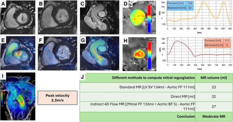
This paper describes the role of cardiovascular magnetic resonance (CMR) imaging in assessing patients with mitral valve disease. Mitral regurgitation (MR) is one of the most prevalent valvular heart diseases. It often progresses without significant symptoms, leading to left ventricular overload, dysfunction, frequent decompensated heart failure episodes, and excess mortality. Cardiovascular magnetic resonance assessment is recommended for MR when routine ultrasound imaging information is insufficient or discordant. A well-planned CMR can provide an in-depth assessment of the mitral valve apparatus, leaflet morphology, and papillary muscles. In addition, it can precisely inform the impact of MR on left atrial and ventricular remodelling. The review aims to highlight established and emerging techniques for morphological assessment, flow assessment (including regurgitation and stenosis), myocardial assessment, and haemodynamic assessment of mitral valve disease by CMR. It also proposes a simplified clinical flow chart for CMR assessment of the mitral valve.
Keywords: Chordae tendineae; Haemodynamics; Heart failure; Heart valve diseases; Humans; Magnetic resonance imaging; Mitral valve; Mitral valve insufficiency; Papillary muscles.
© The Author(s) 2024. Published by Oxford University Press on behalf of the European Society of Cardiology.
Figures


Similar articles
-
Assessment of mitral valve regurgitation by cardiovascular magnetic resonance imaging.Nat Rev Cardiol. 2020 May;17(5):298-312. doi: 10.1038/s41569-019-0305-z. Epub 2019 Dec 9. Nat Rev Cardiol. 2020. PMID: 31819230 Free PMC article. Review.
-
Cardiac magnetic resonance assessment of mitral regurgitation severity appears better than echocardiographic imaging.Int J Cardiovasc Imaging. 2020 May;36(5):889-897. doi: 10.1007/s10554-020-01772-1. Epub 2020 Feb 3. Int J Cardiovasc Imaging. 2020. PMID: 32016882
-
Assessing Regurgitation Severity, Adverse Remodeling, and Fibrosis with CMR in Primary Mitral Regurgitation.Curr Cardiol Rep. 2024 Jul;26(7):705-715. doi: 10.1007/s11886-024-02069-8. Epub 2024 May 15. Curr Cardiol Rep. 2024. PMID: 38748329 Review.
-
Functional valve assessment: the emerging role of cardiovascular magnetic resonance.Methodist Debakey Cardiovasc J. 2010 Jan-Mar;6(1):15-9. doi: 10.14797/mdcj-6-1-15. Methodist Debakey Cardiovasc J. 2010. PMID: 20360653 Review.
-
Disparities in quantification of mitral valve regurgitation between cardiovascular magnetic resonance imaging and trans-thoracic echocardiography: a systematic review.Int J Cardiovasc Imaging. 2025 Apr;41(4):647-658. doi: 10.1007/s10554-024-03280-y. Epub 2024 Nov 5. Int J Cardiovasc Imaging. 2025. PMID: 39499451 Free PMC article.
Cited by
-
Reproducibility and reliability of flow quantification using CMR 2D-phase contrast and 4D-Flow in secondary mitral valve regurgitation.Int J Cardiovasc Imaging. 2025 Jul;41(7):1341-1350. doi: 10.1007/s10554-025-03421-x. Epub 2025 May 16. Int J Cardiovasc Imaging. 2025. PMID: 40377789 Free PMC article.
-
Diagnosis and Diagnostic Challenges of Secondary Mitral Regurgitation in the Era of Transcatheter Edge-to-Edge Repair of the Mitral Valve.J Clin Med. 2025 Jun 26;14(13):4518. doi: 10.3390/jcm14134518. J Clin Med. 2025. PMID: 40648892 Free PMC article. Review.
-
In Vivo Dynamic Coronary Arteries Blood Flow Imaging Based on Multi-Cycle Phase Clustering Ultrafast Ultrasound.Adv Sci (Weinh). 2025 Aug;12(31):e05485. doi: 10.1002/advs.202505485. Epub 2025 Jun 30. Adv Sci (Weinh). 2025. PMID: 40588810 Free PMC article.
References
-
- Otto CM, Nishimura RA, Bonow RO, Carabello BA, Erwin JP, Gentile F, et al. . 2020 ACC/AHA guideline for the management of patients with valvular heart disease: executive summary: a report of the American College of Cardiology/American Heart Association Joint Committee on Clinical Practice Guidelines. Circulation 2021;143:e35–71. 10.1161/CIR.0000000000000932 - DOI - PubMed
Publication types
MeSH terms
LinkOut - more resources
Full Text Sources
