Case Reports
. 2024 Dec:125:110630.
doi: 10.1016/j.ijscr.2024.110630. Epub 2024 Nov 19.

Inguinal swelling unveiling biphasic synovial sarcoma: A case report

Affiliations
Case Reports

Inguinal swelling unveiling biphasic synovial sarcoma: A case report

Faten Limaiem et al. Int J Surg Case Rep. 2024 Dec.

Abstract

Introduction and importance: Synovial sarcoma is a high-grade soft tissue sarcoma primarily affecting teenagers and young adults. Inguinal region involvement is rare, making diagnosis and treatment challenging.

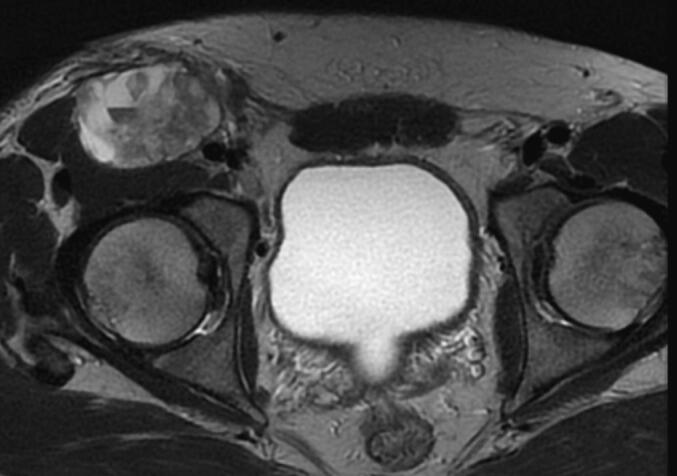
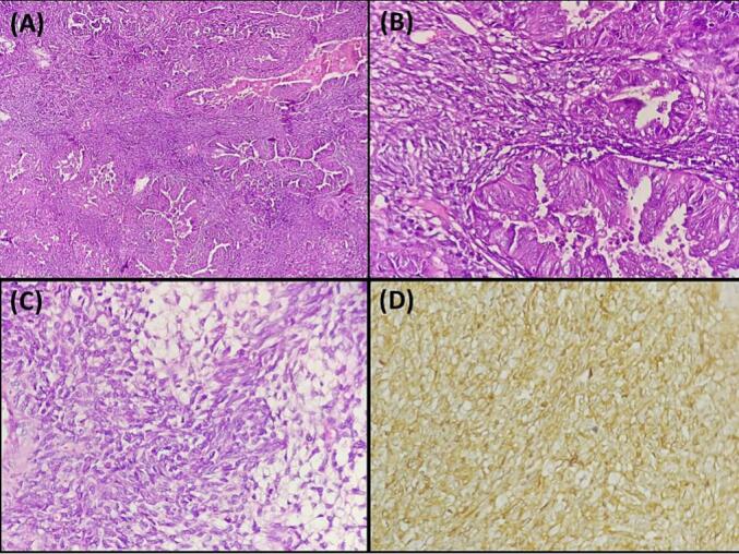
Case presentation: A 35-year-old Tunisian man presented with a progressively enlarging right inguinal swelling. Imaging revealed a mass behind the inguinal ligament, confirmed as biphasic synovial sarcoma through biopsy. The patient underwent successful surgery with clear resection margins. Histopathological examination revealed a biphasic sarcoma with spindle cell and glandular components, supporting the diagnosis of synovial sarcoma. Following surgery, the patient received adjuvant radiotherapy. Regular outpatient follow-up is being conducted to monitor progress.

Clinical discussion: Synovial sarcoma is characterized by slow growth and local invasiveness, with potential for metastasis. It typically presents as a solid mass that can compress nearby structures such as blood vessels. Imaging studies offer valuable insights into tumor location, size, invasiveness, and potential metastases. Local tumor staging relies on MRI, while distant metastases are detected using chest CT or bone scans. Diagnosis is confirmed through histopathological examination and immunohistochemical analysis.

Conclusions: This case report highlights a rare presentation of inguinal synovial sarcoma and emphasizes the importance of individualized multimodal therapy in its management.

Keywords: Immunohistochemistry; Inguinal tumor; Pathology; Soft tissue sarcoma; Surgery; Synovial sarcoma.


Conflict of interest statement

Declaration of competing interest None declared.

Figures

Figs. 1 and 2
Figs. 1 and 2
Pelvic MRI displaying a hyperintense mass on T1-weighted imaging developed in the right inguinal region behind the inguinal ligament and the inguinal fascia. It measures 40 mm in antero-posterior diameter, 60 mm in height, and 62 mm in transverse diameter.
Figs. 1 and 2
Figs. 1 and 2
Pelvic MRI displaying a hyperintense mass on T1-weighted imaging developed in the right inguinal region behind the inguinal ligament and the inguinal fascia. It measures 40 mm in antero-posterior diameter, 60 mm in height, and 62 mm in transverse diameter.
Fig. 3
Fig. 3
A: Histopathological image illustrating a biphasic synovial sarcoma: The image depicts a distinct biphasic pattern characterized by the coexistence of mesenchymal and epithelial differentiation within the tumor (hematoxylin and eosin, magnification × 100). B: Biphasic synovial sarcoma: Image shows spindle cell areas with synovial sarcoma nuclear features and closely packed glandular cells (hematoxylin and eosin, magnification × 400). C: Histopathological image of biphasic synovial sarcoma: The image depicts the mesenchymal component of the tumor, characterized by focal myxoid changes (hematoxylin and eosin, magnification × 400). D: Immunohistochemical staining of synovial sarcoma showing CD99 positivity: The image demonstrates brown staining, indicating positive expression of CD99 in the tumor (immunohistochemistry, × 400).

References

    1. Chaker J., Khlas I., Bouassida I., Ben Ayed A., Bousnina M., Jmal A. Inguinal synovial sarcoma revealed by a limb swelling: a case report. Int. J. Surg. Case Rep. 2024;115 - PMC - PubMed
    1. Chen W.C., Wu P.C., Lin C.Y., Tai T.E. Large inguinal synovial sarcoma mimics a vascular lesion: a case report and literature review. Int. J. Surg. Case Rep. 2020;77:333–336. - PMC - PubMed
    1. Sohrabi C., Mathew G., Maria N., Kerwan A., Franchi T., Agha R.A., et al. The SCARE 2023 guideline: updating consensus Surgical CAse REport (SCARE) guidelines. Int. J. Surg. 2023;109(5):1136. - PMC - PubMed
    1. van der Heide H.J. Synovial sarcoma: oncological and functional results. Eur. J. Surg. Oncol. 1998;24:114–119. - PubMed
    1. Naito N. Synovial sarcoma with a large hematoma in the inguinal region. Arch. Orthop. Trauma Surg. 2000;120:533–534. - PubMed

Publication types

LinkOut - more resources