Case Reports
. 2025 Jun 15;64(12):1893-1899.
doi: 10.2169/internalmedicine.2326-23. Epub 2024 Nov 21.

Positive Antiganglioside Antibodies in a Patient with Primary Diffuse Large B-cell Lymphoma of the Central Nervous System

Affiliations
Case Reports

Positive Antiganglioside Antibodies in a Patient with Primary Diffuse Large B-cell Lymphoma of the Central Nervous System

Ryoji Nishi et al. Intern Med. .

Abstract

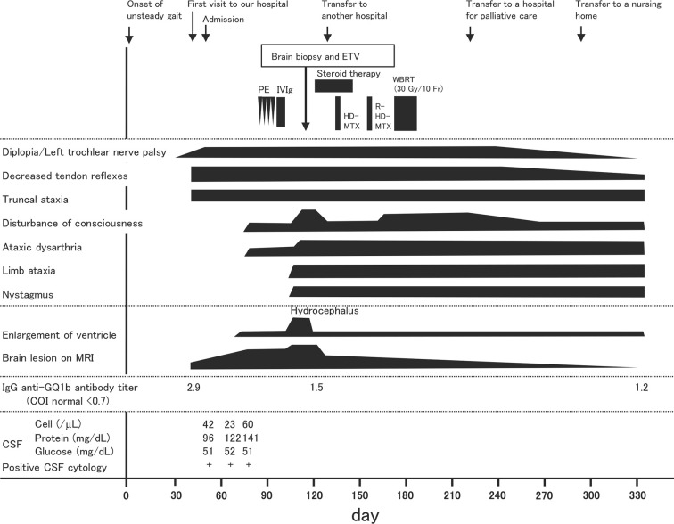
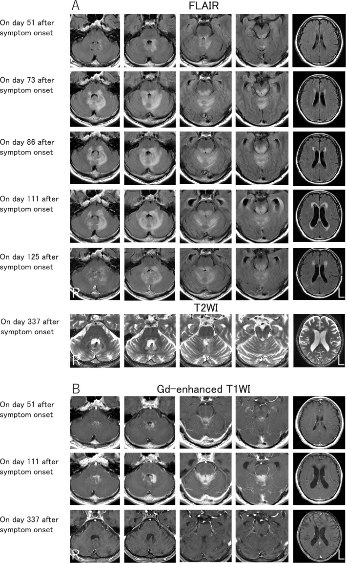
Up to one-third of lymphoma cases involve the nervous system. Miller-Fisher syndrome (MFS) associated with lymphoma is extremely rare. We herein report a case of primary central nervous system lymphoma initially mimicking MFS in a 70-year-old man who presented with subacute unsteady gait and diplopia. A neurological examination revealed unilateral ophthalmoplegia, ataxia, and areflexia. The patient tested positive for anti-GQ1b antibodies, so MFS was initially suspected. However, the progression extended over one month. Subsequently, disturbance of consciousness was observed. Cranial magnetic resonance imaging revealed lesions in the periventricular fourth ventricle, and a brain biopsy indicated diffuse large B-cell lymphoma.

Keywords: Bickerstaff brainstem encephalitis; Miller-Fisher syndrome; anti-GQ1b antibody; anti-GQ1b syndrome; diffuse large B-cell lymphoma; primary central nervous system lymphoma.


Conflict of interest statement

The authors state that they have no Conflict of Interest (COI).

Figures

Figure 1.
Figure 1.
Patient’s clinical course. On admission, the patient manifested diplopia, truncal ataxia, and areflexia. Serum sample was positive for IgG anti-GQ1b antibodies, and brain MRI revealed abnormalities in the brainstem and cerebellum of the periventricular fourth ventricle. A CSF analysis revealed high protein concentrations and cell counts, and a cytological study detected abnormal lymphocytes. PE and IVIg were initiated but were not beneficial. The brain lesions worsened, leading to obstructive hydrocephalus; therefore, ETV was performed. Primary CNS lymphoma disappeared after chemotherapy and WBRT. Positivity for IgG anti-GQ1b antibodies persisted for 11 months after disease onset. CSF: cerebrospinal fluid, CNS: central nervous system, ETV: endoscopic third ventriculostomy, HD-MTX: high-dose methotrexate, IVIg: intravenous immunoglobulin therapy, MRI: magnetic resonance imaging, PE: plasma exchange, R: rituximab, WBRT: whole-brain radiation therapy
Figure 2.
Figure 2.
Brain MRI findings. Representative fluid-attenuated inversion recovery, T2WI (A), and gadolinium-enhanced T1W1 (B) on brain MRI. Contrast-enhanced brain MRI revealed abnormalities in the brainstem and cerebellum of the periventricular fourth ventricle (A and B). The lesions worsened, resulting in obstructive hydrocephalus, which was eventually improved with ETV. Chemotherapy and WBRT were initiated, leading to lymphoma recovery with complete response/unconfirmed results, as shown by enhanced MRI (B). ETV: endoscopic third ventriculostomy, MRI: magnetic resonance imaging, WBRT: whole-brain radiation therapy, T2W1: T2-weighted image, T1W1: T1-weighted image
Figure 3.
Figure 3.
Pathological findings of a brain biopsy specimen. On day 115 after symptom onset, a specimen collected from the cerebellum of the periventricular fourth ventricle was biopsied. A pathological examination revealed proliferation of large, round atypical lymphocytes (A) that were negative for CD3 (C) but positive for CD20 (B) and Ki-67 (D). A brain biopsy indicated diffuse large B-cell lymphoma. Several lymphoma cells were positively stained with anti-GM1 polyclonal antibodies (E); however, none showed clear staining with anti-GQ1b monoclonal antibodies (F). Scale bars=50 μm (A-F).

References

    1. Mauermann ML. Neurologic complications of lymphoma, leukemia, and paraproteinemias. Continuum (Minneap Minn) 23(3, Neurology of Systemic Disease): 669-690, 2017. - PubMed
    1. Giglio P, Gilbert MR. Neurologic complications of non-Hodgkin's lymphoma. Curr Hematol Malig Rep 1: 214-219, 2006. - PubMed
    1. Makranz C, Arkadir D, Nachmias B, et al. Neurological misdiagnoses of lymphoma. Neurol Sci 42: 1933-1940, 2021. - PubMed
    1. Nayak L, Batchelor TT. How I treat neurologic complications in patients with lymphoid cancer. Blood 139: 1469-1478, 2022. - PubMed
    1. Tomita M, Koike H, Kawagashira Y, et al. Clinicopathological features of neuropathy associated with lymphoma. Brain 136: 2563-2578, 2013. - PubMed

Publication types

MeSH terms