. 2024 Nov 20;15(1):9883.
doi: 10.1038/s41467-024-53504-x.

Rotary mechanism of the prokaryotic Vo motor driven by proton motive force

Affiliations

Rotary mechanism of the prokaryotic Vo motor driven by proton motive force

Jun-Ichi Kishikawa et al. Nat Commun. .

Abstract

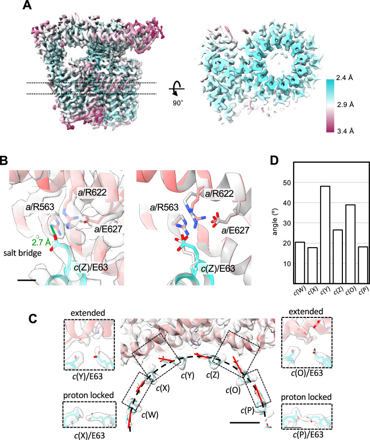
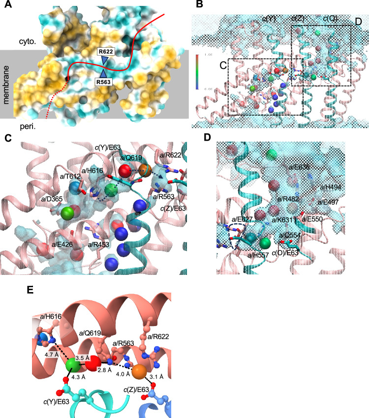
ATP synthases play a crucial role in energy production by utilizing the proton motive force (pmf) across the membrane to rotate their membrane-embedded rotor c-ring, and thus driving ATP synthesis in the hydrophilic catalytic hexamer. However, the mechanism of how pmf converts into c-ring rotation remains unclear. This study presents a 2.8 Å cryo-EM structure of the Vo domain of V/A-ATPase from Thermus thermophilus, revealing precise orientations of glutamate (Glu) residues in the c12-ring. Three Glu residues face a water channel, with one forming a salt bridge with the Arginine in the stator (a/Arg). Molecular dynamics (MD) simulations show that protonation of specific Glu residues triggers unidirectional Brownian motion of the c12-ring towards ATP synthesis. When the key Glu remains unprotonated, the salt bridge persists, blocking rotation. These findings suggest that asymmetry in the protonation of c/Glu residues biases c12-ring movement, facilitating rotation and ATP synthesis.


Conflict of interest statement

Competing interests: The authors declare no competing interests.

Figures

Fig. 1
Fig. 1. Structure of Vo domain in holo-V/A-ATPase from Thermus thermophilus.
A Schematic of V/A-ATPase. The a, c12, and d subunits of the Vo domain are colored pink, blue, and green, respectively. B Schematic of the half-channel model. In T. thermophilus the a subunit of the Vo domain (a-subunit in) forms half-channels on both the periplasmic and cytoplasmic sides of the membrane. H+ can access the c12-ring through these channels, which are separated by two Arg residues. C The atomic model of the Vo domain of holo-V/A-ATPase state 1. The subunits are colored the same as in (A). The scale bar shows 20 Å. D The rotation angles, calculated by UCSF Chimera, between each state are indicated. To help visualize the rotation of the c12-ring, one of the c-subunits is colored blue. The scale bar shows 15 Å. E The interactions between the soluble arm of a-subunit and d-subunit in each state. The density maps are shown semi-transparent. The residues participating in the interactions between subunits are shown in stick representation. The scale bar is 5 Å. F The distance between c/E63 and a/R563, R622 in each state. The values indicate the distances between the Cβ of each residue. The density maps are shown as semi-transparent. The scale bar shows 5 Å.
Fig. 2
Fig. 2. Structure of the isolated Vo domain.
A The density map of the isolated Vo domain. The map is colored according to the local resolution (see key). Left: overall structure. Right: the cross-section image of the region is indicated by the dotted lines in the left panel. B The salt bridge formed between a/R563, a/R622, and c/E63. The residues are shown as sticks. The density maps are shown as semi-transparent. The scale bar shows 3 Å. The left panel represents a comparison of the salt bridge structure of isolated Vo and Vo in the holo-enzyme. The superimposed image of the salt bridge of the isolated Vo (colored) and the Vo domain of holo-enzyme (gray). C The angles adopted by the c/E63 residues. The dashed semi-circle indicates the circumference of the c12-ring. The red lines indicate the angle of the E63 side chain of each c-subunit. The scale bar shows 10 Å. The insets are the magnified views of the regions indicated by the dotted rectangles. The scale bar shows 5 Å. D Plot of the angles of c/E63.
Fig. 3
Fig. 3. Proton pathway in both hemi channels with water densities.
A The surface properties of the transmembrane region of a-subunit. The surface of the a-subunit is colored cyan (hydrophilic) to yellow (hydrophobic). B The coordination of water molecules in the half-channels. The cryo-EM water molecules are shown in sphere representation and colored by density from the MD simulation. The color scale illustrates the relative density of water, with the bulk density set to 1. The iso-surface of the relative water density of 0.5 obtained from MD simulation is shown with the cryo-EM water molecules. The regions indicated by the dashed rectangles are enlarged in (C, D). Enlarged views of the periplasmic (C) and cytoplasmic (D) channels. The residues forming the channels are shown in stick representation. The dashed line in (C) indicates the possible proton relay path. E The possible proton relay path. The cryo-EM water molecules are represented as spheres with the same color coding as (BD).
Fig. 4
Fig. 4. Protonation state-dependent rotation of the c-ring.
A Initial (left) and final structures with protonation (right, upper) and deprotonation (right, lower) states of c/E63 after the 500 ns simulations. The amino acid residues (c/E63, a/R563, and a/Q619) are shown in colored stick representation. The c(Y), c(Z), and c(O) subunits are shown in green, cyan, and blue, respectively. Hydrogen atoms are represented as white sticks. B Rotation angle of the c-ring observed during the simulations. Rotation trajectories in the c(Y)/E63(COOH), c(Z)/E63(COOH), c(O)/E63(COO-) simulations are shown by orange lines, while those in the c(Y)/E63(COOH), c(Z)/E63(COO-), c(O)/E63(COO-) simulations are indicated by blue lines. C The distance between E63 and Q619 during the simulation. The distances obtained in the c(Y)/E63(COOH), c(Z)/E63(COOH), c(O)/E63(COO-) simulations are shown by the orange lines, while those obtained in the c(Y)/E63(COOH), c(Z)/E63(COO-), c(O)/E63(COO-) are indicated by the blue lines. In the MD simulations independently performed three times for 100 ns under the same conditions, a rotation of about 5° in the direction of ATP synthesis was observed when c(Z)/E63 was protonated. Additionally, when protonated, the distance between c(Y)/E63(COOH) and a/Q619 approached that required for hydrogen bond formation, approximately 2 Å. These results were not obtained when c(Z)/E63 was deprotonated (Supplementary Figs. 8, 9).
Fig. 5
Fig. 5. MD simulation of forced rotation of c12-ring.
Snapshot structures obtained during forced-rotation simulation in c(Y)/E63(COOH), c(Z)/E63(COOH), c(O)/E63(COO-) (A), and c(Y)/E63(COOH), c(Z)/E63(COO-), c(O)/E63(COO-) (B). The c(Y), c(Z), and c(O) subunits are shown in green, cyan, and blue, respectively. Three E63 and one T64 from c-subunits, R563 and Q619 from a-subunit are shown in stick representation. The images represent the arrangement of the side chains at 0, 10, and 30 degree rotations. Distances between the salt-bridge c(Z)/E63 –a/R563 and c(O)/E63 - a/R563 are illustrated in cyan and blue lines, respectively, obtained from the c(Y)/E63(COOH), c(Z)/E63(COOH), c(O)/E63(COO-) simulation (C) and the c(Y)/E63(COOH), c(Z)/E63(COO-), c(O)/E63(COO-) simulation (D). Distances of c(Z)/E63 – a/Q619 are also shown in light blue in both figures. E Distances between c(Z)/E63 and c(Y)/T64 (blue line) and between c(Y)/E63 and c(X)/T64 (green line) during the 30° forced c12-ring rotation.
Fig. 6
Fig. 6. Brownian ratchet rotation of c12-ring by pmf.
A A schematic of the interface between the a-subunit and the c12-ring. The ring is surrounded by lipids, as shown here. B is a magnified view of the interface. C The rotation scheme of c12-ring. top: The protonation of c(Z)/E63 causes cleavage of the salt bridge with the Arg residues of the a-subunit. middle: The deprotonated c(O)/E63 is attracted to the Arg residues of the a-subunit by Coulombic interaction (red arrow). The Brownian motion of the protonated (hydrophobic) c(Y)/E63 and c(Z)/E63 is biased toward the membrane (black arrows). bottom: After a 30° rotation, c(O)/E63 forms a new salt bridge with the Arg residues. c(P)/E63 is deprotonated.

Similar articles

Cited by

References

    1. Boyer, P. D. The binding change mechanism for ATP synthase–some probabilities and possibilities. Biochim. Biophys. Acta1140, 215–250 (1993). - PubMed
    1. Mitchell, P. Chemiosmotic coupling in oxidative and photosynthetic phosphorylation. Biol. Rev. Camb. Philos. Soc.41, 445–502 (1966). - PubMed
    1. Senior, A. E. ATP synthesis by oxidative phosphorylation. Physiol. Rev.68, 177–231 (1988). - PubMed
    1. Yoshida, M., Muneyuki, E. & Hisabori, T. ATP synthase - a marvellous rotary engine of the cell. Nat. Rev. Mol. Cell Biol.2, 669–677 (2001). - PubMed
    1. Kuhlbrandt, W. Structure and mechanisms of F-type ATP synthases. Annu. Rev. Biochem.88, 515–549 (2019). - PubMed

Publication types

MeSH terms

LinkOut - more resources