. 2024 Nov 20;13(1):40.
doi: 10.1038/s41389-024-00542-1.

Identification of pancreatic cancer-specific protease substrates for protease-dependent targeted delivery

Affiliations

Identification of pancreatic cancer-specific protease substrates for protease-dependent targeted delivery

Etienne J Slapak et al. Oncogenesis. .

Abstract

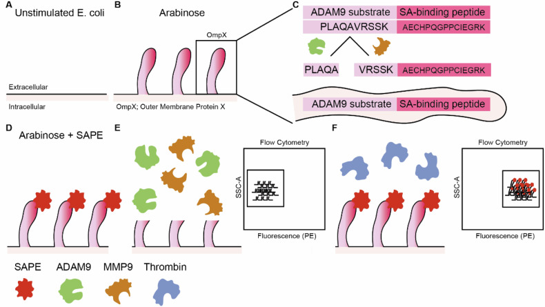
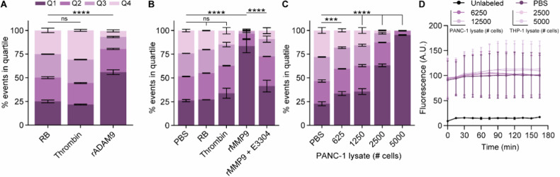
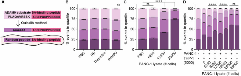
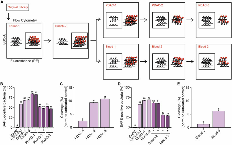
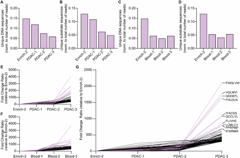
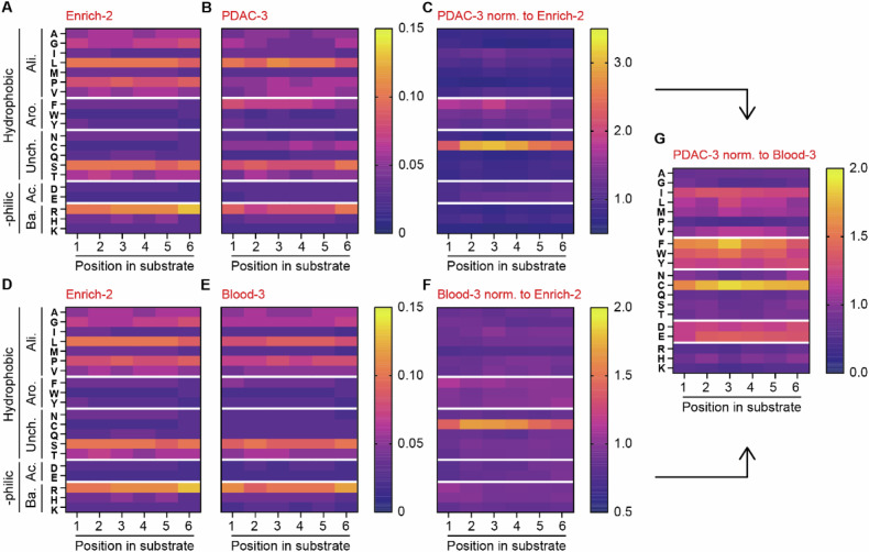
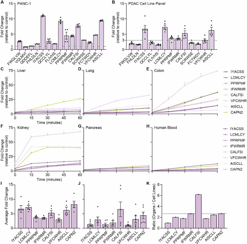
Pancreatic ductal adenocarcinoma (PDAC) presents significant challenges due to the inadequacy of existing chemotherapeutics, which often result in toxicity-dependent dose limitations and premature cessation of therapy. Targeted delivery of therapeutic molecules offers a promising solution. Given that PDAC is marked by a desmoplastic reaction with extensive aberrant protease activity, protease-dependent targeted delivery could minimize off-target toxicities and is of increasing interest. The efficacy of targeted delivery hinges on the specificity of the substrates used; insufficient specificity can lead to off-target effects, reducing the advantage over non-targeted methods. Here, we employ an unbiased library approach to screen over 7 million peptide substrates for proteolytic cleavage by PDAC cell lysates, identifying 37 substrates enriched by at least 500-fold after three rounds of selection. As systemically administered targeted delivery depends on the absence of substrate cleavage in circulation, the peptide library was also screened against whole blood lysates, and enriched substrates were removed from the PDAC-enriched dataset to obtain PDAC-specific substrates. In vitro validation using FRET-peptides showed that 13 of the selected 15 substrates are cleaved by a panel of PDAC cell line lysates. Moreover, evaluation against healthy murine organ and human blood lysates to assess off-target cleavage revealed that the identified substrates are indeed PDAC-specific and that several substrates may be superior with respect to PDAC specificity over the CAPN2-responsive substrate, which has recently shown preclinical potential in targeted therapy, but future animal models should address the potential superiority. Overall, we thus identified substrates with high selectivity and sensitivity for PDAC that could be employed in protease-dependent targeted therapies.


Conflict of interest statement

Competing interests: M.F.B. has received research funding from Celgene, Lead Pharma, and Frame Therapeutics, and acted as a consultant to Servier, Olympus, and Wholomics. None of these were involved in the design of this study nor the drafting of the paper. Ethical approval: The use of whole blood was evaluated and approved by the Ethics Committee of the Amsterdam University Medical Centers and performed following the instructions from the Declaration of Helsinki. The animal tissues were collected in accordance with the regulations on animal experimentation and approved by the ethics committee of Amsterdam University Medical Centers (Approval number: AVD118002015222). Ethics approval and consent to participate: All methods were performed in accordance with the relevant guidelines and regulations. Informed consent: The blood donor has provided consent for the usage of the collected blood in scientific research.

Figures

Fig. 1
Fig. 1. Schematic overview of the CLiPS approach.
A Unstimulated E. coli does not express outer membrane protein X (OmpX) and the rNstretch. B Incubation with arabinose evokes the production of OmpX and outer membrane exposure of rNstretch. C Overview of OmpX containing the ADAM9-substrate and streptavidin-binding sequences. D Incubation of ADAM9 bacteria with streptavidin-coupled phycoerythrin (SAPE) results in fluorescent labeling. E Incubation of fluorescent ADAM9 bacteria with ADAM9 and MMP9 cleaves the substrate with subsequent loss of fluorescent signal, measurable by fluorescence-activated cell sorting (FACS). F Incubation with thrombin, a protease unable to cleave the ADAM9 substrate, has no effect on the fluorescent signal of SAPE-labeled ADAM9 bacteria.
Fig. 2
Fig. 2. ADAM9 bacteria are efficiently cleaved by rADAM9, rMMP9, and PDAC lysates.
The influence of thrombin and rADAM9 (A), rMMP9 alone or in the presence of the MMP inhibitor E3304 (B), and PANC-1 lysate (C) Incubation of ADAM9 bacteria on the percentage of SAPE-positive bacteria. D Fluorescence levels of SAPE-labeled ADAM9 bacteria upon PANC-1 and THP-1 lysate incubation under static conditions as measured by Biotek over time. Data are shown as the mean ± SD of one representative experiment with n = 2, except for (A) and (D), which represent two experiments with n = 2. Two-way ANOVA was used to compare % of events in Q1. Levels of significance: ns not significant, *** p < 0.001, **** p < 0.0001.
Fig. 3
Fig. 3. Complexity of the CLiPS Library is illustrated by differing cleavage efficiencies upon incubation with a single protease as opposed to cell lysates containing various proteases.
A Schematic overview of the generation of the CLiPS Library by replacing the ADAM9-responsive substrate PLAQAVRSSK present in ADAM9 bacteria with a random stretch of 6 amino acids. The effect of thrombin and rMMP9 (B), PANC-1 lysate (C), and combined PANC-1 and THP-1 lysate (D) incubation of the CLiPS Library on the percentage of SAPE-positive bacteria. Data are shown as the mean +/− SD of one representative experiment with n = 2. Two-way ANOVA was used to compare % of events in Q1. Levels of significance: ns = not significant, **** p < 0.0001.
Fig. 4
Fig. 4. Sequential incubation of the CLiPS library with PANC-1 or blood lysates results in the enrichment of SAPE-negative bacteria, indicative of cleaved substrates.
A Schematic overview of the sorting process. B The percentage of SAPE-positive bacteria when untreated (Untreated) SAPE-positive cells are selected (Enrich-1, Enrich-2) and when SAPE-negative bacteria are selected after consecutive incubation with PANC-1 lysate (PDAC-1, PDAC-2, PDAC-3 representing the consecutive rounds of selection). (−). − represents control labeling (PBS), + incubation with PANC-1 lysate. C Normalized reduction of SAPE-positive bacteria after sequential rounds of incubation with PANC-1 lysate (PDAC-1, PDAC-2, PDAC -3 representing the consecutive rounds of selection). D The percentage of SAPE-positive bacteria when untreated SAPE-positive cells are selected (Enrich-1, Enrich-2) and when SAPE-negative bacteria are selected after consecutive incubation with blood lysate (Blood-2, Blood-3). − represents control labeling (PBS), + incubation with blood lysate. E Normalized reduction of SAPE-positive bacteria after sequential rounds of incubation with blood lysate. Blood-1 is missing in panels D and E due to a technical error during data acquisition.
Fig. 5
Fig. 5. CLiPS Library is enriched for PDAC or blood-responsive substrates after incubation with corresponding cell lysates.
A Normalized count of unique DNA sequences after sequential rounds of incubation with PDAC lysate. B Normalized count of unique substrate sequences after sequential rounds of incubation with PDAC lysate. C Normalized count of unique DNA sequences after sequential rounds of incubation with Blood lysate. D Normalized counts of unique substrate sequences after sequential rounds of incubation with Blood lysate. E Fold change of top 100 enriched substrates during sequential incubation with PDAC lysate compared to Enrich-2. F Fold change of top 100 enriched substrates during sequential incubation with Blood lysate compared to Enrich-2. G Fold change of top 100 enriched substrates after removal of all substrates with a fold increase of ≥10 in Blood-3 from PDAC-3. Enrich-2 constitutes the library before the start of the enrichment experiments with PANC1 or blood lysates. Pink lines in figures EG resemble the 10 most enriched hits.
Fig. 6
Fig. 6. PANC-1 and blood lysate show differential amino acid preferences.
A, D Amino acid distribution across substrates in the CLiPS Library at the start of the sorting experiment (Enrich-2). B Amino acid distribution after three incubation rounds with PANC-1 lysate (PDAC-3). C Change in amino acid ratio in PDAC-3 normalized to Enrich-2. E Amino acid distribution after three incubation rounds with blood lysate (Blood-3). F Change in the amino acid ratio in Blood-3 normalized to Enrich-2. G Comparison of amino acid ratio between normalized PDAC-3 and Blood-3 samples. Arrows indicate sample comparison.
Fig. 7
Fig. 7. Validation of the top 15 substrates confirms the identification of PDAC-specific substrates.
A Fold change in fluorescent signal of FRET-peptides after 1 h of incubation with PANC-1 cell lysate versus incubation PBS (control treatment). The dotted line at y = 1 represents the absence of substrate cleavage. Data depict the mean ± SEM of two representative experiments with n = 2. B Fold change in fluorescence of selected substrates upon incubation with lysates from a PDAC cell line panel consisting of BxPC-3, Capan-1, Capan-2, MIA PaCa-2, and PANC-1. The dotted line at y = 3 represents cut- off point. Data depict the mean ± SEM of two representative experiments with n = 2. Cleavage kinetics of substrates with an average of >3-fold change in PDAC panel, and CAPN2-responsive substrate upon incubation with murine liver (C), lung (D), colon (E), kidney (F), pancreas (G), and human whole blood (H) cell lysate. Data are shown as the mean ± SEM of one representative experiment with n = 2. Average fold change of selected peptides after PDAC cell line panel (I) and organ (J) incubation. Data are shown as the mean ± SEM of two representative experiments with n = 2. All data are normalized to corresponding PBS-treated FRET-peptides. K Normalization of the average fold change of organ incubation to average fold change of PDAC cell line incubation. The dotted line represents the ratio of CAPN2-responsive substrate.

Similar articles

References

    1. Kappelhoff R, Puente XS, Wilson CH, Seth A, López-Otín C, Overall CM. Overview of transcriptomic analysis of all human proteases, non-proteolytic homologs and inhibitors: organ, tissue and ovarian cancer cell line expression profiling of the human protease degradome by the CLIP-CHIP™ DNA microarray. Biochim Biophys Acta Mol Cell Res. 2017;1864:2210–9. - PubMed
    1. Doucet A, Butler GS, Rodríguez D, Prudova A, Overall CM. Metadegradomics: toward in vivo quantitative degradomics of proteolytic post-translational modifications of the cancer proteome. Mol Cell Proteom. 2008;7:1925–51. - PubMed
    1. Patel S. A critical review on serine protease: key immune manipulator and pathology mediator. Allergol Immunopathol. 2017;45:579–91. - PMC - PubMed
    1. Ceuleers H, Van Spaendonk H, Hanning N, Heirbaut J, Lambeir AM, Joossens J, et al. Visceral hypersensitivity in inflammatory bowel diseases and irritable bowel syndrome: the role of proteases. World J Gastroenterol. 2016;22:10275–86. - PMC - PubMed
    1. Walsh PN, Ahmad SS. Proteases in blood clotting. Essays Biochem. 2002;38:95–111. - PubMed

LinkOut - more resources