. 2024 Nov 20;14(1):28745.
doi: 10.1038/s41598-024-79024-8.

Serum protein and imaging biomarkers after intermittent steroid treatment in muscular dystrophy

Affiliations

Serum protein and imaging biomarkers after intermittent steroid treatment in muscular dystrophy

Alexander B Willis et al. Sci Rep. .

Abstract

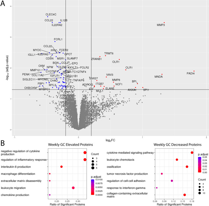
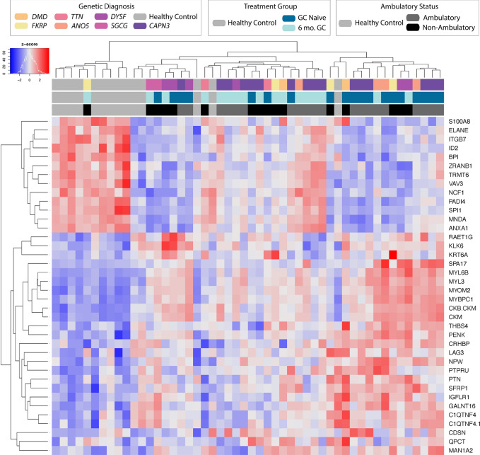
Weekly Steroids in Muscular Dystrophy (WSiMD) was a pilot study to evaluate once weekly prednisone in patients with Limb Girdle and Becker muscular dystrophy (LGMD and BMD, respectively). At study endpoint, there were trends towards increased lean mass, reduced fat mass, reduced creatine kinase and improved motor function. The investigation was motivated by studies in mouse muscular dystrophy models in which once weekly glucocorticoid exposure enhanced muscle strength and reduced fibrosis. WSiMD participants provided blood samples for aptamer serum profiling at baseline and after 6 months of weekly steroids. A subset completed magnetic resonance (MR) evaluation of muscle at study onset and endpoint. At baseline compared to age and sex-matched healthy controls, the aggregate serum protein profile in the WSiMD cohort was dominated by muscle proteins, reflecting leak of muscle proteins into serum. Disease status produced more proteins differentially present in serum compared to steroid-treatment effect. Nonetheless, a response to prednisone was discernable in the WSiMD cohort, even at this low dose. Glucocorticoids decreased muscle proteins and increased certain immune process- and matrix-associated proteins. Muscle MR fat fraction showed trends with functional status. The prednisone-responsive markers could be used in larger trial of prednisone efficacy.

Keywords: Glucocorticoid; Limb girdle muscular dystrophy; MRI; Muscle; Muscular dystrophy; Prednisone; Serum biomarkers.


Conflict of interest statement

Declarations. Competing interests: EMM has been a consultant to Amgen, AstraZeneca, Cytokinetics, Pfizer, Tenaya Therapeutics, and is a founder of Ikaika Therapeutics. ARD is CSO of Ikaika Therapeutics. The other authors have declared that no conflict of interest exists. There are no nonfinancial conflicts to report.

Figures

Fig. 1
Fig. 1
MD Associated Biomarkers. (A) Volcano plot of aptamer profiling from WSiMD participants compared to age/sex matched controls compared using a linear mixed effects model. Benjamini–Hochberg corrected p-value < 0.05 and abs(log2FC) > 0.5 were used as thresholds for significance. (B) Gene Ontology terms were used to cluster the increased (upper panel) and decreased (lower panel) groupings of protein biomarkers. Increased proteins were largely proteins important for muscle structure, function, and development. Decreased proteins included those implicated in cellular adhesion, membrane fusion, actin organization, growth factor signaling and coagulation.
Fig. 2
Fig. 2
Serum protein markers clustered by disease status and treatment group. Heatmap depicting the behavior of the most significant (p < 0.00001) biomarkers associated with LGMD.
Fig. 3
Fig. 3
Protein biomarkers in WSiMD participants responsive to 6 months of intermittent glucocorticoid (GC) treatment. (A) Volcano plot depicting results from linear mixed-effects model for prednisone treatment status. Benjamini–Hochberg corrected p-value < 0.05 and abs(log2FC) > 0.2 were used as thresholds for significance. (B) Gene Ontology groups of GC increased and GC decreased serum proteins. Increased serum proteins are implicated in extracellular matrix disassembly and immune/inflammatory function. Decreased serum proteins were largely extracellular matrix proteins, proteins involved in muscle structure and function, growth factor signaling, and immune/inflammatory cell adhesion and chemotaxis.
Fig. 4
Fig. 4
Individual shifts in prednisone- and disease-associated Biomarkers. Boxplots with paired results depicting representative behavior of biomarkers which had a significant response in both LGMD and the paired prednisone comparison. P-values are derived from the linear mixed model, thresholds are (* p < 0.05, ** p < 0.01, *** p < 0.001, **** p < 0.0001). (A) Glucocorticoid-only responsive marker. (B) Clinically monitored creatine kinase and troponin markers. (C) Proteins displaying both dystrophy association and glucocorticoid responsiveness. GC treatment had disease normalizing effects in this group.
Fig. 5
Fig. 5
Muscular Dystrophy-associated, GC responsive signature separates control serum profiles from GC treated from GC naïve sera. Heatmap depicting the behavior of muscular dystrophy associated, glucocorticoid responsive serum biomarkers.
Fig. 6
Fig. 6
MRI and biomarkers related to functional outcomes. (A) Muscle fat fraction at study onset and after 6 months of weekly prednisone (V1, visit 1; V2, visit 2) in the quadriceps (RED) and Triceps surae (BLUE) muscle groups. Participants are listed by gene name along the X axis. Data was available from two muscle groups for 10 participants. For the two FKRP participants and one ANO5 participant, data was only available from the Triceps surae (blue). The majority of participants experienced a small increase in muscle fat fraction in the quadriceps and Triceps surae muscle groups. There was not always agreement between the two muscles. (B) Spearman correlation coefficients between age, functional performance measures, MRI water T2 and muscle fat fraction, and body composition as determined by DEXA scanning. (C) Example biomarkers showing a significant association (FDR ≤ 0.05) by linear regression with NSAD measure. The muscle protein markers CKM and PGAM2 correlated positively with functional performance as assessed by NSAD. D) Broad trends towards CKM and CCL33 reduction with change in NSAD over six months of treatment with once weekly prednisone (p value ≤ 0.05, FDR ≥ 0.05).

Update of

References

    1. Bushby, K. et al. Diagnosis and management of Duchenne muscular dystrophy, Part 1: Diagnosis, and pharmacological and psychosocial management. Lancet Neurol.9, 77–93 (2010). - PubMed
    1. Fenichel, G. M. et al. Long-term benefit from prednisone therapy in duchenne muscular dystrophy. Neurology41, 1874–1877 (1991). - PubMed
    1. Guglieri, M. et al. Effect of different corticosteroid dosing regimens on clinical outcomes in boys with Duchenne muscular dystrophy: A randomized clinical trial. Jama327, 1456–1468 (2022). - PMC - PubMed
    1. Schara, U., Mortier & Mortier, W. Long-term steroid therapy in Duchenne muscular dystrophy-positive results versus side effects. J. Clin. Neuromuscul. Dis.2, 179–183 (2001). - PubMed
    1. Weber, D. R., Hadjiyannakis, S., McMillan, H. J., Noritz, G. & Ward, L. M. Obesity and endocrine management of the patient with Duchenne muscular dystrophy. Pediatrics142, S43-s52 (2018). - PMC - PubMed

MeSH terms

LinkOut - more resources