

Optimized simple culture protocol for inducing mature myotubes from MYOD1-overexpressed human iPS cells
- PMID: 39567611
- PMCID: PMC11579357
- DOI: 10.1038/s41598-024-79745-w
Optimized simple culture protocol for inducing mature myotubes from MYOD1-overexpressed human iPS cells
Abstract
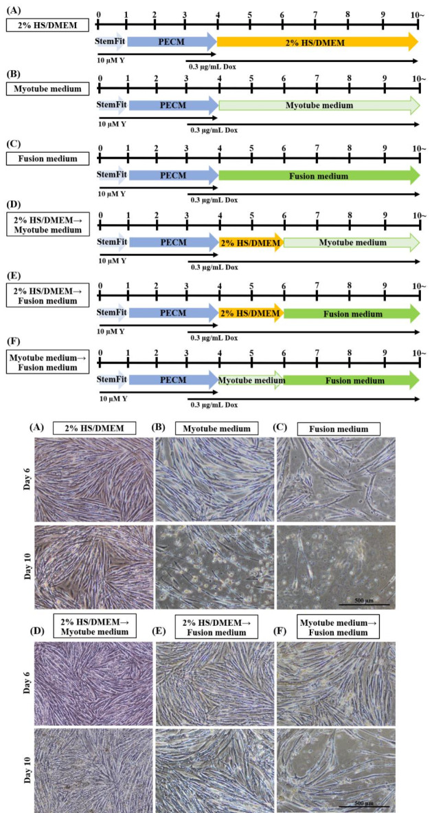
The forced expression system of MYOD1, a master gene for myogenic differentiation, can efficiently and rapidly reproduce muscle differentiation of human induced pluripotent stem cells (hiPSCs). Despite these advantages of the MYOD1 overexpression system, developed myotubes are relatively immature and do not recapitulate several aspects of striated muscle fibers. Here, we developed a simple optimized protocol using an alternative culture medium for maximizing the advantages of the MYOD1 overexpression system, and successfully improved the formation of multinucleated mature myotubes within 10 days. In this study, we generated hiPSCs derived from healthy donors and an individual with congenial muscular dystrophy caused by LMNA mutation (laminopathy), and compared disease-associated phenotypes in differentiated myotubes generated by the conventional method and by our new optimized culture method. Using our optimized method, abnormal myonuclear shape was pronounced in the patient-derived iPSCs. In addition, abnormal accumulation of the nuclear membrane protein emerin was observed in LMNA-mutant hiPSCs. Our new culture method is expected to be widely applicable as a MYOD1 overexpression model of hiPSC-derived skeletal muscle cells for the analysis of a variety of muscle diseases.
Keywords: Differentiation of skeletal muscle cells; Induced pluripotent stem cells; Laminopathy; Muscular dystrophy; Newly developed culture method.
© 2024. The Author(s).
Conflict of interest statement
Declarations. Competing interests: The authors declare no competing interests.
Figures


Similar articles
-
Efficient and reproducible myogenic differentiation from human iPS cells: prospects for modeling Miyoshi Myopathy in vitro.PLoS One. 2013 Apr 23;8(4):e61540. doi: 10.1371/journal.pone.0061540. Print 2013. PLoS One. 2013. PMID: 23626698 Free PMC article.
-
Myogenic differentiation of muscular dystrophy-specific induced pluripotent stem cells for use in drug discovery.Stem Cells Transl Med. 2014 Feb;3(2):149-60. doi: 10.5966/sctm.2013-0095. Epub 2014 Jan 6. Stem Cells Transl Med. 2014. PMID: 24396035 Free PMC article.
-
Directed Myogenic Differentiation of Human Induced Pluripotent Stem Cells.Methods Mol Biol. 2016;1353:89-99. doi: 10.1007/7651_2015_257. Methods Mol Biol. 2016. PMID: 25971915
-
Induction of Skeletal Muscle Progenitors and Stem Cells from human induced Pluripotent Stem Cells.J Neuromuscul Dis. 2020;7(4):395-405. doi: 10.3233/JND-200497. J Neuromuscul Dis. 2020. PMID: 32538862 Free PMC article. Review.
-
Recent advances in animal and human pluripotent stem cell modeling of cardiac laminopathy.Stem Cell Res Ther. 2016 Sep 20;7(1):139. doi: 10.1186/s13287-016-0401-5. Stem Cell Res Ther. 2016. PMID: 27649756 Free PMC article. Review.
Cited by
-
Generation of bovine iPSCs from fetal fibroblasts for in vitro myogenesis and cultured meat.Front Nutr. 2025 May 16;12:1562981. doi: 10.3389/fnut.2025.1562981. eCollection 2025. Front Nutr. 2025. PMID: 40453727 Free PMC article.
-
Early and non-destructive prediction of the differentiation efficiency of human induced pluripotent stem cells using imaging and machine learning.Sci Rep. 2025 Jul 23;15(1):26821. doi: 10.1038/s41598-025-11108-5. Sci Rep. 2025. PMID: 40702033 Free PMC article.
References
-
- Takahashi, K. & Yamanaka, S. Induction of pluripotent stem cells from mouse embryonic and adult fibroblast cultures by defined factors. Cell126, 663–676. 10.1016/j.cell.2006.07.024 (2006). - PubMed
-
- Takahashi, K. et al. Induction of pluripotent stem cells from adult human fibroblasts by defined factors. Cell131, 861–872. 10.1016/j.cell.2007.11.019 (2007). - PubMed
MeSH terms
Substances
Grants and funding
- 2022-2024/Follow-up Grant from Tokyo Medical University
- JP17bm0804005/Acceleration Program for Intractable Disease Research Utilizing Disease-specific iPS cells
- 20H03594/KAKENHI
- 23ek0109639h0001/Practical Research Project for Rare/Intractable Diseases
- 29-4/Intramural Research Grant for Neurological and Psychiatric Disorders of NCNP
LinkOut - more resources
Full Text Sources
Miscellaneous
