

Distribution and evidence of co-infection of the two microsporidian parasites Astathelohania contejeani and Nosema austropotamobii in Austropotamobius pallipes complex in Northern and Central Italy
- PMID: 39567840
- PMCID: PMC12038494
- DOI: 10.1017/S0031182024001525
Distribution and evidence of co-infection of the two microsporidian parasites Astathelohania contejeani and Nosema austropotamobii in Austropotamobius pallipes complex in Northern and Central Italy
Abstract
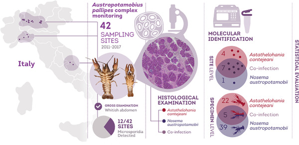
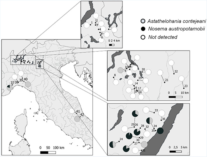
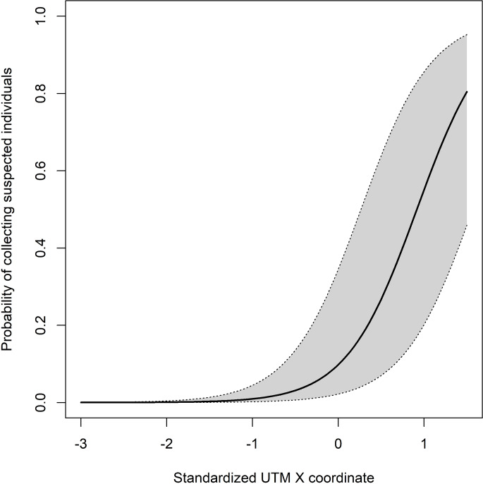
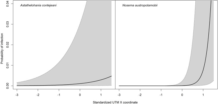
Austropotamobius pallipes complex is an endangered freshwater crayfish species in Europe and the assessment of the health status of its wild populations is essential for conservation purposes. The two microsporidia Astathelohania contejeani and Nosema austropotamobii have been reported to cause in A. pallipes complex a chronic parasitic infection, known as ‘porcelain disease’, which reduces population fitness and leads the host to death. Due to the similar macroscopic signs produced, molecular biology analyses are required to unambiguously distinguish between these microsporidia. Focusing on A. pallipes complex populations located in Northern and Central Italy, the present study provides an evaluation of prevalence and distribution of A. contejeani and N. austropotamobii, and investigates the variables affecting the probability of detecting infected specimens during a survey (e.g. sex, crayfish density, longitude). Microsporidia were identified in 12 populations among the 42 monitored from 2011 to 2017, with an average prevalence of 3.12% for A. contejeani and 3.60% for N. austropotamobii, the latter being reported in a wider area than previously documented (from Lombardy to Liguria Regions). Notably, crayfish co-infected by both microsporidia were also detected in 4 populations. Moreover, it was observed that the probability of detecting a crayfish with a microsporidian infection significantly increased eastwards in the studied area, especially for N. austropotamobii. Our distribution map for microsporidiosis, combined with molecular screening, will be useful for planning breeding and translocation efforts for A. pallipes complex populations.
Keywords: detection probability; parasite; porcelain disease; white-clawed crayfish.
Conflict of interest statement
The authors declare there are no conflicts of interest. The funders had no role in the study's design, data collection, analysis, or interpretation, as well as in manuscript writing, or the decision to publish the results.
Figures
Similar articles
-
Ultrastructural and molecular characterization of Vairimorpha austropotamobii sp. nov. (Microsporidia: Burenellidae) and Thelohania contejeani (Microsporidia: Thelohaniidae), two parasites of the white-clawed crayfish, Austropotamobius pallipes complex (Decapoda: Astacidae).J Invertebr Pathol. 2018 Jan;151:59-75. doi: 10.1016/j.jip.2017.11.002. Epub 2017 Nov 6. J Invertebr Pathol. 2018. PMID: 29122615
-
Horizontal transmission of Thelohania contejeani in the endangered white-clawed (Austropotamobius pallipes) and the invasive signal crayfish (Pacifastacus leniusculus).Parasitology. 2012 Sep;139(11):1471-7. doi: 10.1017/S0031182012000777. Epub 2012 Jul 20. Parasitology. 2012. PMID: 22814084
-
Patterns of infection in a native and an invasive crayfish across the UK.J Invertebr Pathol. 2021 Sep;184:107595. doi: 10.1016/j.jip.2021.107595. Epub 2021 Apr 18. J Invertebr Pathol. 2021. PMID: 33878331
-
Nosema bombycis: A remarkable unicellular parasite infecting insects.J Eukaryot Microbiol. 2024 Sep-Oct;71(5):e13045. doi: 10.1111/jeu.13045. Epub 2024 Aug 2. J Eukaryot Microbiol. 2024. PMID: 39095558 Review.
-
The Crayfish Plague Pathogen Aphanomyces astaci in Ireland.Microorganisms. 2024 Jan 4;12(1):102. doi: 10.3390/microorganisms12010102. Microorganisms. 2024. PMID: 38257929 Free PMC article. Review.
Cited by
-
Better Alone Than in Bad Company: Trophic Ecology of Co-Occurring Invasive and Native Crayfish.Ecol Evol. 2025 May 14;15(5):e71385. doi: 10.1002/ece3.71385. eCollection 2025 May. Ecol Evol. 2025. PMID: 40370355 Free PMC article.
References
-
- Anderson TK and Sukhdeo MVK (2013) The relationship between community species richness and the richness of the parasite community in Fundulus heteroclitus. Journal of Parasitology 99, 391–396. - PubMed
-
- Anderson LG, Bojko J, Bateman KS, Stebbing PD, Stentiford GD and Dunn AM (2021) Patterns of infection in a native and an invasive crayfish across the UK. Journal of Invertebrate Pathology 184, 107595. - PubMed
-
- Azevedo C (1987) Fine structure of the microsporidan Abelspora portucalensis gen.n., sp.n. (Microsporida) parasite of the hepatopancreas of Carcinus maenas (Crustacea, Decapoda). Journal of Invertebrate Pathology 49, 83–92.
-
- Bates D, Mächler M, Bolker BM and Walker SC (2015) Fitting linear mixed-effects models using lme4. Journal of Statistical Software 67, 1–48.
MeSH terms
LinkOut - more resources
Full Text Sources
