

Evaluation of root canal morphology of mandibular premolars in Pakistani population using the new classification: a CBCT study
- PMID: 39568006
- PMCID: PMC11577896
- DOI: 10.1186/s12903-024-05149-x
Evaluation of root canal morphology of mandibular premolars in Pakistani population using the new classification: a CBCT study
Abstract
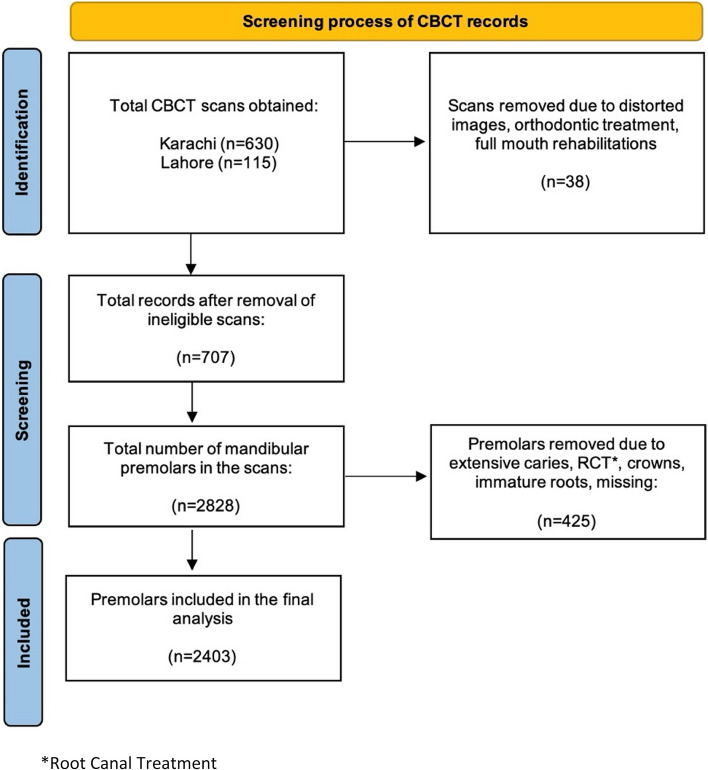
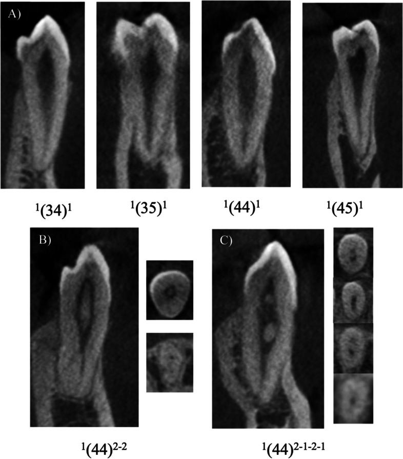
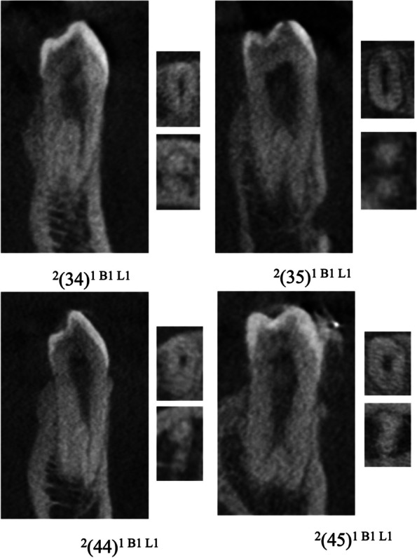
Background: A comprehensive understanding of the root form and canal anatomy is essential for successful endodontic treatment. This study aimed to evaluate the root canal anatomy of mandibular premolars in the Pakistani population using cone beam computed tomography (CBCT) and to classify the findings with the new classification proposed by Ahmed et al. METHODS: Ethical exemption was obtained from Aga Khan University Hospital, Karachi. A total of 707 CBCT scans from Karachi and Lahore were included, comprising 592 scans from a tertiary care hospital in Karachi and 115 scans from a radiology center in Lahore. The study focused on sound, fully formed mandibular first and second premolars, excluding those with significant caries, restorations, or prior root canal treatments. Scans from different equipment were used, and calibration was achieved between a specialist endodontist and two dental residents. Data was analyzed using the Statistical Package for the Social Sciences (SPSS) software, version 26. Descriptive statistics, Chi-square tests to determine association between the variables, and a significance level set at 5% (p <0.05) were utilized.
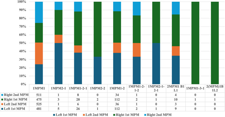
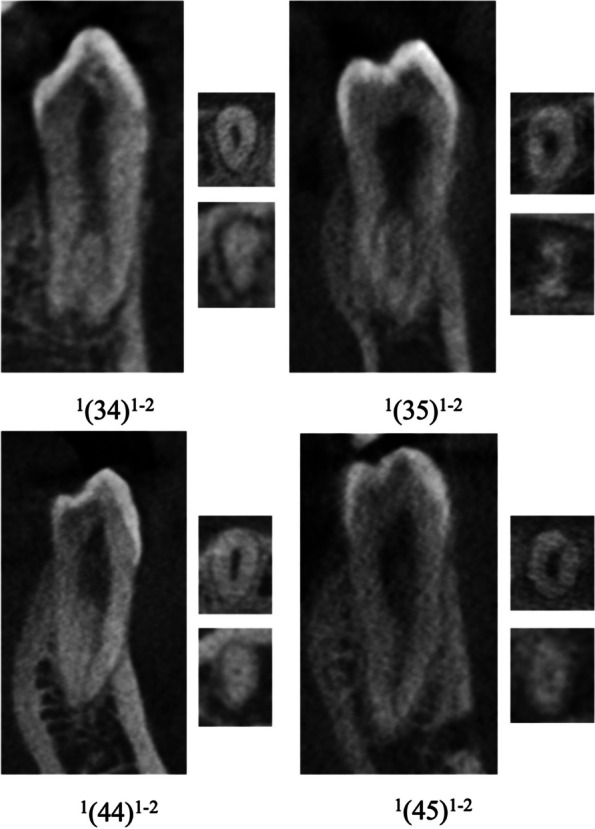
Results: A total of 2403 mandibular premolars were analyzed. The most common configuration was 1MPM1 (82.90%), with rare variations such as 1MPM1-3-1 (0.04%) and 2MPM1 B1 L2 (0.04%). Single-rooted premolars were predominant (98.87%), and no significant differences were observed when data was stratified by age or gender.
Conclusions: This is the first study in Pakistan involving multiple centers and using the classification system by Ahmed et al. to understand the anatomy of mandibular premolars. The findings indicate that while most premolars have a single root and canal, variations exist. These variations highlight the importance of understanding canal morphology for improving the success of endodontic treatment. Future studies should include a larger and more diverse dataset to fully represent the Pakistani population.
Keywords: CBCT; Mandibular premolars; Root canal anatomy; Root canal variations.
© 2024. The Author(s).
Conflict of interest statement
Declarations. Ethics approval and consent to participate: This study has been conducted in accordance with the guidelines of the Declaration of Helsinki. Additionally, Ethical exemption (reference no. 2024–10008-29535) was obtained from the Ethical Review Committee at the Aga Khan University before initiating the study. The Ethical Review Committee of Aga Khan University, Karachi waived the informed consent procedure because the study is retrospective in nature. Consent for publication: Not applicable. Competing interests: The authors declare no competing interests.
Figures


Similar articles
-
Comprehensive analysis of root canal morphology in maxillary premolars among the Pakistani subpopulation: a CBCT-based study.Eur J Med Res. 2024 Jul 27;29(1):391. doi: 10.1186/s40001-024-01990-6. Eur J Med Res. 2024. PMID: 39068434 Free PMC article.
-
Investigating root and canal morphology of anterior and premolar teeth using CBCT with a novel coding classification system in Saudi subpopulation.Sci Rep. 2025 Feb 5;15(1):4392. doi: 10.1038/s41598-025-86277-4. Sci Rep. 2025. PMID: 39910098 Free PMC article.
-
Evaluation of root and canal morphology of mandibular premolar amongst Saudi subpopulation using the new system of classification: a CBCT study.BMC Oral Health. 2023 May 15;23(1):291. doi: 10.1186/s12903-023-03002-1. BMC Oral Health. 2023. PMID: 37189077 Free PMC article.
-
Micro computed tomography (Micro-CT) characterization of root and root canal morphology of mandibular first premolars: a systematic review and meta-analysis.BMC Oral Health. 2024 Jan 2;24(1):1. doi: 10.1186/s12903-023-03624-5. BMC Oral Health. 2024. PMID: 38167114 Free PMC article.
-
Systematic review and meta-analysis of root morphology and canal configuration of permanent premolars using cone-beam computed tomography.BMC Oral Health. 2024 Jun 4;24(1):656. doi: 10.1186/s12903-024-04419-y. BMC Oral Health. 2024. PMID: 38835024 Free PMC article.
References
-
- Habib AA, Kalaji MN, Al saysd TJ, Al jawfi KA. Root canal configurations of the first and second mandibular premolars in the population of north Syria. Journal of Taibah University Medical Sciences. 2015;10(4):391–5.
-
- Cleghorn BM, Christie WH, Dong CC. The root and root canal morphology of the human mandibular first premolar: a literature review. J Endod. 2007;33(5):509–16. - PubMed
-
- Cleghorn BM, Christie WH, Dong CC. The root and root canal morphology of the human mandibular second premolar: a literature review. J Endod. 2007;33(9):1031–7. - PubMed
MeSH terms
LinkOut - more resources
Full Text Sources
Miscellaneous
