. 2025 Mar 1;110(3):725-738.
doi: 10.3324/haematol.2024.286418.

Targeting P-selectin and interleukin-1β in mice with sickle cell disease: effects on vaso-occlusion, liver injury and organ iron deposition

Affiliations

Targeting P-selectin and interleukin-1β in mice with sickle cell disease: effects on vaso-occlusion, liver injury and organ iron deposition

Érica M F Gotardo et al. Haematologica. .

Abstract

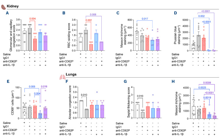
Continuous vaso-occlusive and inflammatory processes cause extensive end-organ damage in adults with sickle cell disease (SCD), and there is little evidence that long-term hydroxyurea therapy prevents this. In initial trials, P-selectin blockade with crizanlizumab reduced SCD vaso-occlusive crisis frequency, and interleukin (IL)-1β inhibition in SCD patients, using canakinumab, lowered inflammatory markers. We used murine SCD models to examine the effects of acute and chronic blockade of P-selectin and of IL-1β on vaso-occlusive events, their inflammatory profile and organ health. Both approaches improved impaired cutaneous microvascular perfusion in SCD mice by reducing TNF-α-induced vaso-occlusion. Acute P-selectin blockade markedly reduced TNF-α-induced neutrophil-platelet aggregate formation in SCD mice, and decreased leukocyte- rolling movements in the microvasculature, while acute IL-1β inhibition attenuated microvascular leukocyte adhesion. Six weeks of IL-1β-blocking immunotherapy improved the inflammatory profile of SCD mice, considerably reduced hepatic fibrosis and provided some relief from lung injury. In contrast, although P-selectin blockade reduced glomerular congestion, no significant benefit to overall organ pathology was observed. Unexpectedly, while combining the two immunotherapies reduced microvascular occlusion, their prolonged use caused acute liver injury. Notably, inhibition of IL-1β, but not of P-selectin, remarkably decreased hemosiderosis, in association with reduced tissue macrophage infiltration and the correction of biomarkers of dysregulated iron turnover. Our findings suggest that the attenuation of inflammation, as well as of vaso- occlusive processes, may be crucial for mitigating organ damage in SCD. Future trials should explore the ability of cytokine blockade to prevent multi-organ damage in patients with SCD, beyond evaluating vaso-occlusive crisis frequency.


Figures

Figure 1.
Figure 1.
Acute P-selectin blockade and IL-1b neutralization in mice with sickle cell disease reverse TNF-α cytokine-induced cutaneous microvascular dysfunction, in association with differing effects on heterocellular aggregation formation. (A) Experimental design for acute protocols in sickle cell disease (SCD) mice. Townes SCD mice were treated intraperitoneally (i.p.) with antibodies targeting P-selectin (anti-CD62P) and/or IL-1b (anti-IL-1b), at 3.5 hours (h) or 24 h (respectively) prior to analysis, consisting of laser Doppler flowmetry of the skin, blood sampling or intravital microscopy. Mice were also subjected to laser Doppler flowmetry at 48 h before protocols to measure basal hemodynamics. Doses were administered as follows: saline (100 μL, 24 h before analysis, N=5-6); 30 μg/mouse IgG1 isotype control antibody (A110-1, 3.5 h before analysis, N=6); 30 μg/mouse anti-CD62P (RB40.34, 3.5 h before analysis, N=5-6); 200 μg/mouse anti-IL-1b (01BSUR, 24 h before analysis, N=5-6), and 30 μg/mouse anti-CD62P plus 200 μg/mouse anti-IL-1b (N=6). Inflammatory processes were induced in mice by the administration of TNF-a (TNF) at 3 h before flowmetry/blood sampling/intravital microscopy (0.5 μg, i.p.). (B) Basal levels of IL-1b were measured in the serum of C57BL/6J mice (C57, N=10) and Townes SCD mice (Tow, N=13) by enzyme-linked immunosorbant assay. (C) Flow cytometry was used to monitor neutrophil-platelet heterocellular aggregate formation in the blood of SCD mice. Platelet-neutrophil aggregates were determined as the percentage of CD45+Ly6G+ neutrophils in peripheral blood samples of mice that also labeled positive for the platelet marker, CD41. Laser Doppler flowmetry (PeriFlux 6000; Perimed) was used to measure blood flow (D) and perfusion (E) in the hindlimb superficial microcirculation of SCD mice. Data were normalized relative to basal measurements taken from the same mice, 48 h earlier. (B-E) Statistical comparisons are made between treatments and their mechanistic control, and between treatments.
Figure 2.
Figure 2.
Acute P-selectin blockade and IL-1b neutralization modulate TNFα-induced leukocyte recruitment and vaso-occlusive-like processes in mice with sickle cell disease. Townes sickle cell disease (SCD) mice were treated intraperitoneally (i.p.) with antibodies targeting P-selectin (anti-CD62P) and/or the inflammasome-processed cytokine, IL-1b (anti-IL-1b), at 3.5 or 24 hours (h), respectively, prior to intravital microscopy (N=5-6 per group) (see Figure 1A). Vaso-occlusive processes were induced in mice by the administration of TNF-α (TNF, 0.5 μg, i.p.): leukocyte (white blood cell [WBC]) recruitment was quantified in 5-9 venules of the cremaster muscle of each mouse at 180 minutes (min) after TNF. Graphs depict WBC rolling along the venule walls (100 μm) (A), WBC adhesion (B), and WBC extravasation (C). Circles represent values for each venule. (D) Representative photomicrographs from a cremaster venule in each treatment group; bars represent 20 µm. Note the visible improvement in venular blood flow in animals treated with anti-CD62P, anti-IL-1b, or their combination, compared to the venules of TNF-challenged mice (and IgG1/TNF-treated mice), where individual cells (indicated by *) can be distinctly observed, indicating vaso-occlusion. White arrows indicate the direction of blood flow. Serum concentrations of (E) soluble ICAM-1 (sICAM-1), and (F) IL-10 were determined by enzyme-linked immunosorbant assay. (A-C) and (E-F), statistical comparisons are made between treatments and their mechanistic control, and between treatments.
Figure 3.
Figure 3.
Anti-inflammatory effects of chronic administration (6 weeks) of immunotherapies that block P-selectin and/or IL-1b in sickle cell disease mice. (A) Experimental design: Berkeley sickle cell disease (SCD) mice (4-months old) were treated with intraperitoneal (i.p.) injections of 30 μg/mouse anti-CD62P (N=9-10) or equivalent IgG1 (N=9), three times a week, and/or 7.5 mg/ kg of anti-IL1b antibody (N=7-9) or saline (N=10), twice a week, for 6 weeks. At 48 hours (h) after administration of the final intervention dose, peripheral blood samples and organs of mice were collected and appropriately stored for processing. (B) Basal levels of IL-1b were measured in the serum of C57BL/6J mice (C57, N=10) and Berkeley SCD mice (Berk, N=7) by enzyme-linked immunosorbant assay (ELISA). (C) Platelet-neutrophil aggregates were monitored in peripheral blood samples by flow cytometry, and serum concentrations of (D) TNF-α, (E) IL-6 and (F) IFN-y were determined by ELISA. Platelet-neutrophil aggregates are expressed as the percentage of neutrophils (CD45+/Ly6G+) that also displayed labeling for the platelet marker, CD41. (C-F) Statistical comparisons are made between treatments and their mechanistic control, and between treatments.
Figure 4.
Figure 4.
Effects of 6-weeks administration of anti-CD62P and anti-IL-1b immunotherapies on liver histology, injury, and iron accumulation in sickle cell disease mice. Berkeley sickle cell disease (SCD) mice (4-months old) were treated with immunotherapies, or not, as described in Figure 3A, for 6 weeks. At 48 hours (h) after administration of the final intervention dose, livers were dissected and processed for histological analysis (hematoxylin and eosin [H&E] staining, 20x magnification [mag.]), analysis of fibrosis (Masson Trichrome, 20x mag.), iron accumulation (Perls Prussian blue, 63x mag.), and CD68+ cells (anti-CD68 immunohistochemical staining, 20x mag.). Image depicts representative photographs of stained sections from each treatment group and comparison with liver sections from hemizygous Berkeley control mice of the same age. Photographs are representative of 4 images per section (2-4 sections per mouse). Black arrows: congested vessels; yellow arrows: collagen deposits; yellow circles: areas of macrophage infiltration. Photomicrographs taken with a Zeiss Axio Imager D2.
Figure 5.
Figure 5.
Effects of 6-weeks administration of anti-CD62P and anti-IL-1b immunotherapies on kidney histology, injury, and iron accumulation in sickle cell disease mice. Berkeley sickle cell disease (SCD) mice (4-months old) were treated, or not, with immunotherapies, as described in Figure 3A, for 6 weeks. At 48 hours (h) after administration of the final intervention dose, kidneys were dissected and processed for histological analysis (hematoxylin and eosin [H&E] staining, 20x magnification [mag.]), analysis of fibrosis (Masson Trichrome, 20x mag.), iron accumulation (Perls Prussian blue, 63x mag.), and CD68+ cell infiltration (anti-CD68 immunohistochemical staining, 20x mag.). Image depicts representative photographs of stained sections from each treatment group and comparison with kidney sections from hemizygous Berkeley control mice of the same age. Photographs are representative of 4 images per section (2-4 sections per mouse). Black arrows: congested vessels; yellow arrows: collagen deposits; yellow circles: areas of macrophage infiltration. Photomicrographs taken with a Zeiss Axio Imager D2.
Figure 6.
Figure 6.
Morphometric analysis, histopathological evaluation and quantification of collagen, iron and macrophage infiltration in the livers of Berkeley sickle cell disease mice after 6 weeks of immunotherapies. Berkeley sickle cell disease (SCD) mice (4-months old) were treated, or not, with immunotherapies, as described in Figure 3A, for 6 weeks. At 48 hours (h) after administration of the final intervention dose, livers were dissected and processed for analysis in 2-4 histological slices per animal (N=4/ group). The following analyses were performed in hematoxylin and eosin-stained sections: (A) morphometric analysis of the number of congested vessels (count µm-2); (B) congestion in sinusoids and vessels; (C) hepatic degradation score; (D) centrilobular necrosis score. (E) Analysis of collagen fiber deposition (Masson Trichrome; AU staining intensity µm-2). (F) Hemosiderin quantification (Perls Prussian blue staining; staining intensity µm-2), and quantification of (G) monocyte/macrophage infiltration (anti-CD68 immunohistochemical staining; AU of staining intensity µm-2).
Figure 7.
Figure 7.
Histopathological evaluation and quantification of collagen, iron and macrophage infiltration in the kidneys and lungs of Berkeley sickle cell disease mice after 6 weeks of immunotherapies. Berkeley sickle cell disease (SCD) mice (4-months old) were treated, or not, with immunotherapies, as described in Figure 3A, for 6 weeks. At 48 hours (h) after administration of the final intervention dose, kidneys, and lungs were dissected and processed for analysis in 2-4 histological slices per animal (N=4/ group). Kidneys: (A) congestion in glomerular vessels and capillaries; (B) red blood cell (RBC) sickling score. (C) Analysis of collagen fiber deposition (Masson Trichrome; AU staining intensity µm-2). (D) Hemosiderin quantification (Perls Prussian blue; staining intensity µm-2). (E) Analysis of monocyte/ macrophage infiltration (anti-CD68 immunohistochemical staining; AU of staining intensity µm-2). Lungs, hematoxylin and eosin staining: (F) congestion in alveolar vessels; (G) septal thickening score. (H) Analysis of collagen fiber deposition, mainly in the interalveolar septa (Masson Trichrome; AU staining intensity µm-2). Statistical comparisons are made between treatments and their mechanistic control, and between treatments.
Figure 8.
Figure 8.
Effects of 6-weeks administration of anti-CD62P and anti-IL-1b immunotherapies on liver iron content and the gene expression of markers of iron regulation in livers from sickle cell disease mice. Berkeley sickle cell disease (SCD) mice (4-months old) were treated, or not, with immunotherapies, as described in Figure 3A, for 6 weeks. At 48 hours (h) after administration of the final intervention dose, livers were dissected and processed for iron measurement and analysis of gene expression by quantitative polymerase chain reaction (PCR). (A) Total and free iron in liver homogenates (nMols/mg tissue). (B-D) Organs from aged-matched hemizygous littermate mice (Hemi) were used for histopathological comparisons. Expressions of the genes encoding (B) hepcidin (Hamp), (C) bone morphogenetic protein-6 (BMP-6, Bmp6) and (D) transferrin receptor-1 (TrF-1, Tfrc) were normalized to the expression of Actb and Gapdh. Statistical comparisons are made between treatments and their mechanistic control, and between treatments.

Similar articles

References

    1. Kato GJ, Piel FB, Reid CD, et al. . Sickle cell disease. Nat Rev Dis Primers. 2018;4:18010. - PubMed
    1. Rees DC, Williams TN, Gladwin MT. Sickle-cell disease. Lancet. 2010;376(9757):2018-2031. - PubMed
    1. Conran N, Belcher JD. Inflammation in sickle cell disease. Clin Hemorheol Microcirc. 2018;68(2-3):263-299. - PMC - PubMed
    1. Asare K, Gee BE, Stiles JK, et al. . Plasma interleukin-1beta concentration is associated with stroke in sickle cell disease. Cytokine. 2010;49(1):39-44. - PMC - PubMed
    1. Qari MH, Dier U, Mousa SA. Biomarkers of inflammation, growth factor, and coagulation activation in patients with sickle cell disease. Clin Appl Thromb Hemost. 2012;18(2):195-200. - PubMed

MeSH terms

LinkOut - more resources