

Evolution of g-type lysozymes in metazoa: insights into immunity and digestive adaptations
- PMID: 39568508
- PMCID: PMC11576321
- DOI: 10.3389/fcell.2024.1487920
Evolution of g-type lysozymes in metazoa: insights into immunity and digestive adaptations
Abstract
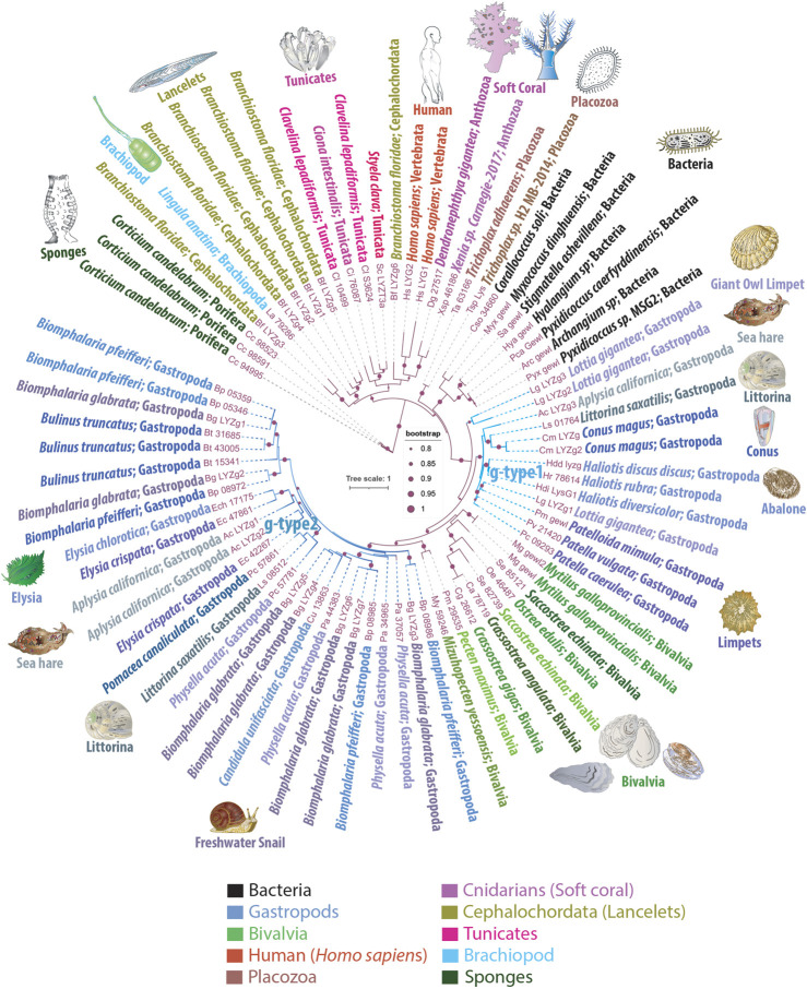
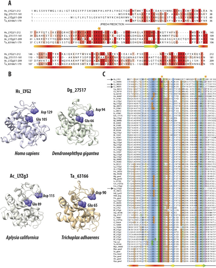
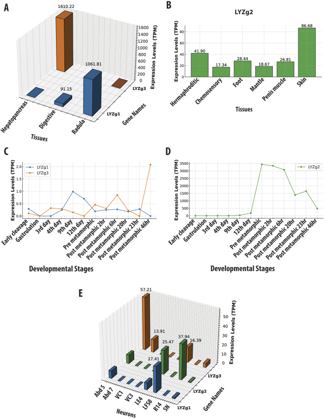
Exploring the evolutionary dynamics of lysozymes is critical for advancing our knowledge of adaptations in immune and digestive systems. Here, we characterize the distribution of a unique class of lysozymes known as g-type, which hydrolyze key components of bacterial cell walls. Notably, ctenophores, and choanoflagellates (the sister group of Metazoa), lack g-type lysozymes. We reveal a mosaic distribution of these genes, particularly within lophotrochozoans/spiralians, suggesting the horizontal gene transfer events from predatory myxobacteria played a role in their acquisition, enabling specialized dietary and defensive adaptations. We further identify two major groups of g-type lysozymes based on their widespread distribution in gastropods. Despite their sequence diversity, these lysozymes maintain conserved structural integrity that is crucial for enzymatic activity, underscoring independent evolutionary pathways where g-type lysozymes have developed functionalities typically associated with different lysozyme types in other species. Specifically, using Aplysia californica as a reference species, we identified three distinct g-type lysozyme genes: two are expressed in organs linked to both feeding and defense, and the third exhibits broader distribution, likely associated with immune functions. These findings advance our understanding of the evolutionary dynamics shaping the recruitment and mosaic functional diversification of these enzymes across metazoans, offering new insights into ecological physiology and physiological evolution as emerging fields.
Keywords: Aplysia californica; ctenophores; gene retention; horizontal gene transfer; innate immunity; placozoa; porifera; spiralia.
Copyright © 2024 Mukherjee and Moroz.
Conflict of interest statement
The authors declare that the research was conducted in the absence of any commercial or financial relationships that could be construed as a potential conflict of interest. The author(s) declared that they were an editorial board member of Frontiers, at the time of submission. This had no impact on the peer review process and the final decision.
Figures
Similar articles
-
A new lysozyme from the eastern oyster, Crassostrea virginica, and a possible evolutionary pathway for i-type lysozymes in bivalves from host defense to digestion.BMC Evol Biol. 2010 Jul 15;10:213. doi: 10.1186/1471-2148-10-213. BMC Evol Biol. 2010. PMID: 20633278 Free PMC article.
-
Identification and expression analysis of the g-type and c-type lysozymes in grass carp Ctenopharyngodon idellus.Dev Comp Immunol. 2010 May;34(5):501-9. doi: 10.1016/j.dci.2009.12.009. Epub 2009 Dec 27. Dev Comp Immunol. 2010. PMID: 20034515
-
Molecular characterization and expression analysis of the chicken-type and goose-type lysozymes from totoaba (Totoaba macdonaldi).Dev Comp Immunol. 2020 Dec;113:103807. doi: 10.1016/j.dci.2020.103807. Epub 2020 Jul 29. Dev Comp Immunol. 2020. PMID: 32735961
-
Lysozymes in the animal kingdom.J Biosci. 2010 Mar;35(1):127-60. doi: 10.1007/s12038-010-0015-5. J Biosci. 2010. PMID: 20413917 Review.
-
Animal lysozymes c and g: an overview.EXS. 1996;75:9-31. doi: 10.1007/978-3-0348-9225-4_2. EXS. 1996. PMID: 8765292 Review.
Cited by
-
The ancestral architecture of the immune system in simplest animals.Front Immunol. 2025 Jan 7;15:1529836. doi: 10.3389/fimmu.2024.1529836. eCollection 2024. Front Immunol. 2025. PMID: 39840034 Free PMC article. No abstract available.
References
-
- Bathige S. D., Umasuthan N., Whang I., Lim B. S., Jung H. B., Lee J. (2013). Evidences for the involvement of an invertebrate goose-type lysozyme in disk abalone immunity: cloning, expression analysis and antimicrobial activity. Fish. Shellfish Immunol. 35 (5), 1369–1379. 10.1016/j.fsi.2013.07.048 - DOI - PubMed
LinkOut - more resources
Full Text Sources