Review
. 2024 Nov 20;5(12):e70010.
doi: 10.1002/mco2.70010. eCollection 2024 Dec.

Ferroptosis: mechanisms and therapeutic targets

Affiliations
Review

Ferroptosis: mechanisms and therapeutic targets

Qian Zhou et al. MedComm (2020). .

Abstract

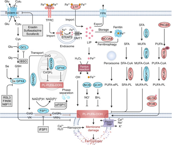
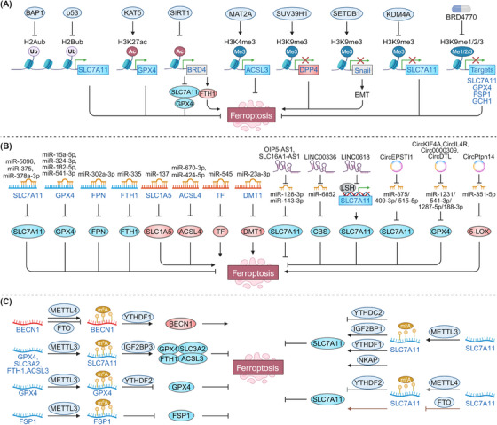
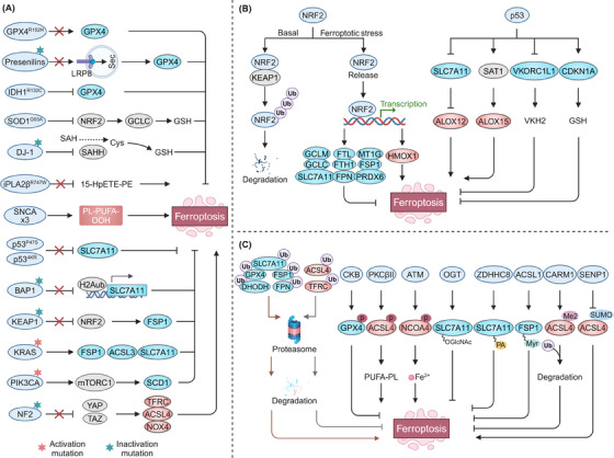
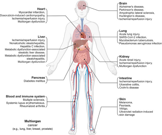
Ferroptosis is a nonapoptotic form of cell death characterized by iron-dependent lipid peroxidation in membrane phospholipids. Since its identification in 2012, extensive research has unveiled its involvement in the pathophysiology of numerous diseases, including cancers, neurodegenerative disorders, organ injuries, infectious diseases, autoimmune conditions, metabolic disorders, and skin diseases. Oxidizable lipids, overload iron, and compromised antioxidant systems are known as critical prerequisites for driving overwhelming lipid peroxidation, ultimately leading to plasma membrane rupture and ferroptotic cell death. However, the precise regulatory networks governing ferroptosis and ferroptosis-targeted therapy in these diseases remain largely undefined, hindering the development of pharmacological agonists and antagonists. In this review, we first elucidate core mechanisms of ferroptosis and summarize its epigenetic modifications (e.g., histone modifications, DNA methylation, noncoding RNAs, and N6-methyladenosine modification) and nonepigenetic modifications (e.g., genetic mutations, transcriptional regulation, and posttranslational modifications). We then discuss the association between ferroptosis and disease pathogenesis and explore therapeutic approaches for targeting ferroptosis. We also introduce potential clinical monitoring strategies for ferroptosis. Finally, we put forward several unresolved issues in which progress is needed to better understand ferroptosis. We hope this review will offer promise for the clinical application of ferroptosis-targeted therapies in the context of human health and disease.

Keywords: epigenetics; ferroptosis; human disease; lipid peroxidation.


Conflict of interest statement

The authors have declared that no conflict of interest exists.

Figures

FIGURE 1
FIGURE 1
Core mechanisms of ferroptosis. Oxidizable lipids in the cellular membrane, particularly PUFA‐PLs mediated by ACSL4 and LPCAT3, are preferential substrates of iron‐dependent nonenzymatic and enzymatic lipid peroxidation. When GPX4‐dependent or ‐independent antioxidant systems (e.g., FSP1, DHODH, GCH1, iNOS, 7‐DHC) are compromised, cellular defense against lipid peroxidation diminishes, allowing uncontrolled lipid peroxidation. The lethal accumulation of lipid peroxidation overwhelms antioxidant defenses and membrane repair capacity, activating mechanosensitive cation channels, disrupting ion homeostasis, and ultimately leading to membrane rupture and ferroptotic cell death. ACSL4, acyl‐CoA synthetase long‐chain family member 4; ALOX, arachidonate lipoxygenase; BH4, tetrahydrobiopterin; BSO, buthionine sulfoximine; CoQ, coenzyme Q; Cys, cysteine; Cys2, cystine; DHODH, dihydroorotate dehydrogenase; DMT1, divalent metal transporter 1; FPN, ferroportin; FSP1, ferroptosis suppressor protein 1; GCH1, GTP cyclohydrolase‐1; GCL, glutamate–cysteine ligase; Glu, glutamate; GPX4, glutathione peroxidase 4; GSH, glutathione; H₂O₂, hydrogen peroxide; LIP, labile iron pool; iNOS, inducible nitric oxide synthase; LPCAT3, lysophosphatidylcholine acyltransferase 3; MBOAT, membrane bound O‐acyltransferase; MUFA, monounsaturated fatty acid; LTF, lactotransferrin; NCOA4, nuclear receptor coactivator 4; PKCβII, protein kinase C; POR, cytochrome P450 oxidoreductase; PUFA, polyunsaturated fatty acid; SCD1, stearoyl‐CoA desaturase; Se, selenium; SFA, saturated fatty acid; STARD7, StAR‐related lipid transfer domain‐containing 7; STEAP3, six‐transmembrane epithelial antigens of the prostate 3; TF, transferrin; TFRC, transferrin receptor; TRP, transient receptor potential; VK, vitamin K.
FIGURE 2
FIGURE 2
Epigenetic regulation in ferroptosis. (A) Posttranslational modifications of histones, such as acylation, methylation, and ubiquitination, regulate DNA accessibility and the expression of ferroptosis‐related genes, thereby modulating the cellular ferroptosis response. (B) miRNAs regulate ferroptosis by inhibiting mRNA translation or promoting mRNA degradation, while lncRNAs and circRNAs function as competing endogenous RNAs (ceRNAs), sponging miRNAs to modulate the expression of ferroptosis‐related genes such as SLC7A11 and GPX4. (C) m6A modifications regulate ferroptosis by altering the mRNA stability of key genes such as SLC7A11, GPX4, and FSP1 through the coordinated actions of methyltransferases, demethylases, and reader proteins. ACSL4, acyl‐CoA synthetase long‐chain family member 4; BAP1, BRCA1‐associated deubiquitinase 1; BECN1, beclin 1; CBS, cystathionine beta‐synthase; DMT1, divalent metal transporter 1; DPP4, dipeptidyl peptidase 4; FPN, ferroportin; FSP1, ferroptosis suppressor protein 1; FTH1, ferritin heavy chain 1; FTO, FTO alpha‐ketoglutarate dependent dioxygenase; GCH1, GTP cyclohydrolase‐1; GPX4, glutathione peroxidase 4; H2Aub, ubiquitination of histones H2A; H2Bub, ubiquitination of histones H2B; IGF2BP3, insulin‐like growth factor 2 mRNA binding protein 3; KAT5, lysine acetyltransferase 5; LSH, lymphoid‐specific helicase; NKAP, NF‐κB activating protein; m6A, N6‐methyladenosine; MAT2A, methionine adenosyltransferase 2A; METTL4, methyltransferase‐like 4; SIRT1, sirtuin 1; SLC1A5, solute carrier family 1 member 5; SLC3A2, solute carrier family 3 member 2; SLC7A11, solute carrier family 7 member 11; Snail, snail family transcriptional repressor 1; TF, transferrin; YTHDF1, YTH N6‐methyladenosine RNA binding protein F1.
FIGURE 3
FIGURE 3
Nonepigenetic regulation in ferroptosis. (A) Genetic mutations in neurodegenerative diseases and cancers are key modulators of pathways influencing ferroptosis susceptibility as shown. (B) NRF2 transcriptionally regulates genes involved in GSH and GPX4 biosynthesis, iron metabolism, NADPH production, and FSP1, thereby modulating cellular susceptibility to ferroptosis. TP53 transcriptionally inhibits SLC7A11 and VKORC1L1 and upregulates SAT1, sensitizing cells to ferroptosis. However, under cystine deprivation, TP53 suppresses ferroptosis by promoting CDKN1A expression. (C) Core ferroptosis‐regulating proteins, including SLC7A11, GPX4, ACSL4, FSP1, and DHODH, can undergo multiple PTMs, such as ubiquitination, phosphorylation, acetylation, O‐GlcNAcylation, S‐palmitoylation, N‐myristoylation, methylation, and SUMOylation, thereby influencing ferroptosis sensitivity. ACSL4, acyl‐CoA synthetase long‐chain family member 4; ALOX, arachidonate lipoxygenase; BAP1, BRCA1‐associated deubiquitinase 1; CARM1, coactivator‐associated arginine methyltransferase 1; DHODH, dihydroorotate dehydrogenase; EGFR, epidermal growth factor receptor; FPN, ferroportin; FSP1, ferroptosis suppressor protein 1; FTH1, ferritin heavy chain 1; GCH1, GTP cyclohydrolase‐1; GPX4, glutathione peroxidase 4; GSH, glutathione; HMOX1, heme oxygenase 1; IDH1, isocitrate dehydrogenase 1; iPLA2β, phospholipase A2β; KEAP1, kelch‐like ECH‐associated protein 1; NF2, neurofibromin 2; NRF2, nuclear factor erythroid 2‐related factor 2; OGT, O‐linked N‐acetylglucosamine (GlcNAc) transferase; SAHH, s‐adenosylhomocysteine hydrolase; SAT1, spermidine/spermine N1‐acetyltransferase 1; SCD1, stearoyl‐CoA desaturase; SENP1, SUMO‐specific peptidase 1; SOD1, superoxide dismutase 1; TF, transferrin; TFRC, transferrin receptor; VKORC1L1, vitamin K epoxide reductase complex subunit 1‐like 1. GCLC, glutamate–cysteine ligase catalytic subunit; SNCA, synuclein α; PIK3CA, phosphatidylinositol‐4,5‐bisphosphate 3‐kinase catalytic subunit α; YAP, Yes1‐associated transcriptional regulator; TAZ, transcriptional coactivator with PDZ‐binding motif; NOX4, NADPH oxidase 4; CDKN1A, cyclin dependent kinase inhibitor 1A; CKB, creatine kinase B; ATM, ATM serine/threonine kinase; ZDHHC8, zinc finger DHHC‐type containing 8.
FIGURE 4
FIGURE 4
Role of ferroptosis in various diseases across different organs and tissues. Ferroptosis serves as an intrinsic tumor‐suppressive mechanism, with its evasion supporting tumorigenesis and progression. Additionally, ferroptosis activation is implicated in the pathogenesis of multiple neurodegenerative diseases, organ injuries, metabolic dysfunction‐associated steatotic liver disease, and dermatological conditions such as psoriasis, vitiligo, and UV‐induced skin damage. Notably, due to its complex interaction with the immune system, ferroptosis may exert dual effects, particularly in immune and infectious diseases.

References

    1. Green DR. The coming decade of cell death research: five riddles. Cell. 2019;177(5):1094‐1107. - PMC - PubMed
    1. Dixon SJ, Lemberg KM, Lamprecht MR, et al. Ferroptosis: an iron‐dependent form of nonapoptotic cell death. Cell. 2012;149(5):1060‐1072. - PMC - PubMed
    1. Xie Y, Hou W, Song X, et al. Ferroptosis: process and function. Cell Death Differ. 2016;23(3):369‐379. - PMC - PubMed
    1. Dixon SJ, Olzmann JA. The cell biology of ferroptosis. Nat Rev Mol Cell Biol. 2024;25(6):424‐442. - PubMed
    1. Lei G, Zhuang L, Gan B. Targeting ferroptosis as a vulnerability in cancer. Nat Rev Cancer. 2022;22(7):381‐396. - PMC - PubMed

LinkOut - more resources