Review
. 2024 Oct 30;15(46):19225-19246.
doi: 10.1039/d4sc05700g. eCollection 2024 Nov 27.

Time-resolved single-cell transcriptomic sequencing

Affiliations
Review

Time-resolved single-cell transcriptomic sequencing

Xing Xu et al. Chem Sci. .

Abstract

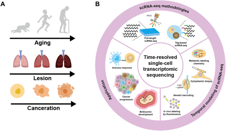
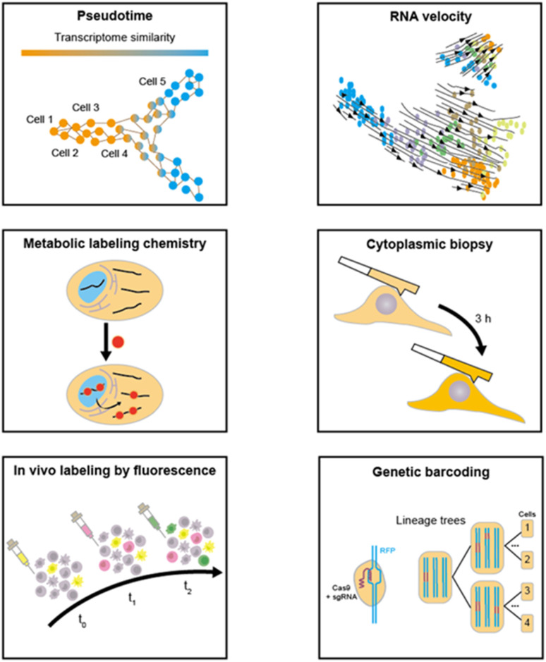
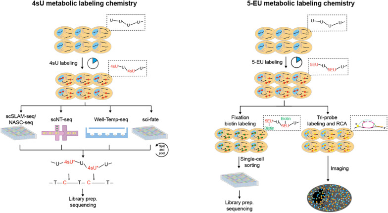
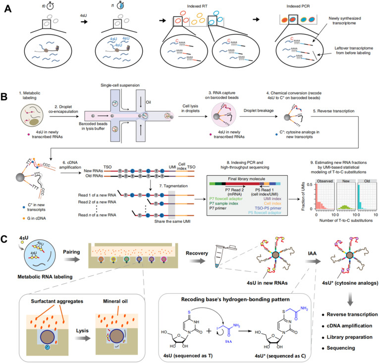
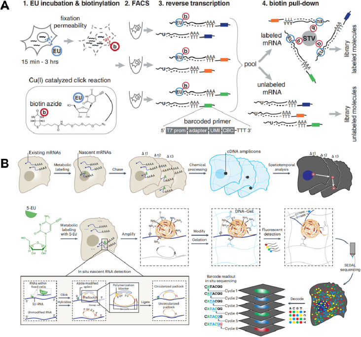
Cells experience continuous transformation under both physiological and pathological circumstances. Single-cell RNA sequencing (scRNA-seq) is competent in disclosing the disparities of cells; nevertheless, it poses challenges in linking the individual cell state at distinct time points. Although computational approaches based on scRNA-seq data have been put forward for trajectory analysis, the result is based on assumptions and fails to reflect the actual states. Consequently, it is necessary to incorporate a "time anchor" into the scRNA-seq library for the temporal documentation of the dynamic expression pattern. This review comprehensively overviews the time-resolved single-cell transcriptomic sequencing methodologies and applications. As scRNA-seq functions as the basis for profiling single-cell expression patterns, the review initially introduces various scRNA-seq approaches. Subsequently, the review focuses on the different experimental strategies for introducing a "time anchor" to scRNA-seq, highlighting their principles, strengths, weaknesses, and comparing their adaptation in various scenarios. Next, it provides a brief summary of applications in immunity response, cancer progression, and embryo development. Finally, the review concludes with a forward-looking perspective on future advancements in time-resolved single-cell transcriptomic sequencing.


Conflict of interest statement

There are no conflicts to declare.

Figures

Fig. 1
Fig. 1. (A) The dynamic alterations in the biological progress at the cell, organ and individual level. (B) Overview of time-resolved single-cell transcriptomic sequencing including approaches for scRNA-seq, strategies for providing temporal modeling of scRNA-seq and the applications for cell dynamics analysis in different fields.
Fig. 2
Fig. 2. Full-length scRNA-seq methods. (A) The flow chart of Smart-seq2 sequencing library preparation. (B) The flow chart of Smart-seq3 sequencing library preparation. (C) The flow chart of VASA-seq sequencing library preparation.
Fig. 3
Fig. 3. Liquid-phase tag based scRNA-seq methods. (A) The flow chart of STRT sequencing library preparation. (B) The flow chart of CEL-seq2 sequencing library preparation. (C) The flow chart of sci-RNA-seq sequencing library preparation.
Fig. 4
Fig. 4. Solid-phase tag based scRNA-seq methods. (A) Schematic workflow of Drop-seq for highly parallel single-cell transcriptome sequencing based on droplet microfluidics. This figure has been reproduced from Cell, 2015, 161, 1202–1214, with permission from Elsevier, copyright 2015. (B) Schematic workflow of Well-Paired-seq that utilizes thousands of microwells for single-cell RNA sequencing. This figure has been reproduced from Small Methods, 2022, 6, e2200341, with permission from Wiley-VCH GmbH, copyright 2022. (C) Schematic workflow of Paired-seq for highly parallel scRNA-seq, which utilizes hydrodynamic traps for single-cell isolation and valve/pump structure for liquid control. This figure has been reproduced from Genome Biol, 2016, 17, 77, with permission from Springer Nature Publishing, copyright 2020. (D) Disco integrates a valve-based strategy with droplet microfluidics for pairing of a single cell and a bead. This figure has been reproduced from Nat Methods, 2022, 19, 323–330, with permission from Springer Nature Publishing, copyright 2022.
Fig. 5
Fig. 5. The overview of various temporal modeling approaches based on scRNA-seq.
Fig. 6
Fig. 6. (A) Chemical structures and abbreviations of nucleoside analogues that have been used for RNA metabolic labelling to incorporate chemical handles. (B) The reaction mechanism of RNA metabolic labelling.
Fig. 7
Fig. 7. The overview of metabolic labeling chemistry in time-resolved scRNA-seq. 4sU and 5-EU are the most prevalently utilized metabolic labels, which can be employed for the detection of nascent RNAs. The metabolic labeling chemistry can be integrated with diverse scRNA-seq methodologies for expression pattern profiling and spatial imaging analysis.
Fig. 8
Fig. 8. (A) Schematic workflow of sci-fate combines sci-RNA-seq with a 4sU metabolic labeling strategy for high-throughput single cell analysis. This figure has been reproduced from Nat Biotechnol, 2020, 38, 980–988, with permission from Springer Nature Publishing, copyright 2020. (B) Schematic workflow of scNT-seq that employs droplet microfluidics for temporally resolved scRNA-seq. This figure has been reproduced from Nat Methods, 2020, 17, 991–1001, with permission from Springer Nature Publishing, copyright 2020. (C) Schematic workflow of Well-Temp-seq that utilizes a microwell device for 4sU-labelled cell loading and pairing. This figure has been reproduced from Nat Commun, 2023, 14, 1272, with permission from Springer Nature Publishing, copyright 2023.
Fig. 9
Fig. 9. (A) Schematic workflow of scEU-seq which integrates 5-EU labeling, click chemistry-based biotinylation, and MARS-seq to simultaneously quantify nascent and pre-existing transcripts in thousands of single cells. This figure has been reproduced from Science, 2020, 367, 1151–1156, with permission from American Association for the Advancement of Science, copyright 2020. (B) Schematic workflow of TEMPOmap that employs 5-EU metabolic labeling and a tri-probe set to spatially and temporally resolve scRNA-seq. This figure has been reproduced from Nat Methods, 2023, 20, 695–705, with permission from Springer Nature Publishing, copyright 2023.
Fig. 10
Fig. 10. Cytoplasmic biopsy based time-resolved scRNA-seq. (A) By sequentially extracting RNAs within a single cell, this approach enables the direct monitoring of cellular dynamics within the same cell. The RNA extracting device includes (B) commercial fluidic force microscopy (FluidFM) used in Live-seq and (C) nanopipette coupled with electrowetting.
Fig. 11
Fig. 11. (A) The strategy of in vivo cell labelling by fluorescence to record different cells at different time points. (B) Schematic workflow of Zman-seq that utilizes fluorescent antibodies at different time points to add temporal information to scRNA-seq data. This figure has been reproduced from Cell, 2024, 187, 149–165 e123, with permission from Elsevier, copyright 2023. (C) Schematic workflow of Gehart's method that leverages cell type-specific reporters for temporal recording and combines scRNA-seq to study time-ordered trajectories during enteroendocrine differentiation. This figure has been reproduced from Cell, 2019, 176, 1158–1173 e1116, with permission from Elsevier, copyright 2019.
Fig. 12
Fig. 12. (A) The schematic illustration of LINNAEUS which utilizes genetic barcodes for constructing the lineage tree of zebrafish line. This figure has been reproduced from Nat Biotechnol, 2018, 36, 469–473, with permission from Springer Nature Publishing, copyright 2018. (B) A universal CRISPR array repair lineage tracing (CARLIN) mouse line constructed by the introduction of Cas9-based scar. The mouse line can be applied in phylogeny analysis, clonal tracing and analysis of functional heterogeneity. This figure has been reproduced from Cell, 2020, 181, 1693–1694, with permission from Elsevier, copyright 2020.
Fig. 13
Fig. 13. Applications of time-resolved scRNA-seq in studying dynamics of the immune response. (A) Zman-seq calculates the value of tumor exposure time and reveals temporal NK cell trajectories in the tumor. The finding also reveals that the TREM2 antagonistic antibody reprograms the tumor microenvironment (TME) by disrupting the transition from monocytes to TAMs. This figure has been reproduced from Cell, 2024, 187, 149–165 e123, with permission from Elsevier, copyright 2024. (B) Schematic diagram for integrating Live-seq with live-cell imaging to detect the immune response of individual macrophage cells. Single RAW264.7 cells are initially subjected to Live-seq and subsequently exposed to LPS while tracking Tnf-mCherry fluorescence through time-lapse imaging. This figure has been reproduced from Nature, 2022, 608, 733–740, with permission from Springer Nature Publishing, copyright 2022.
Fig. 14
Fig. 14. Applications of time-resolved scRNA-seq in studying transcriptional changes of cancer. (A) Schematic illustration of regulon identification through linking TFs with their regulated genes. The heat map presenting the average regulon activity of 3 regulons of HCT116 cells after 5-AZA-CdR treatment. This figure has been reproduced from Nat Methods, 2020, 17, 991–1001, with permission from Springer Nature Publishing, copyright 2023. (B) The subtype switching of single GBM brain tumor cells identified by the cytoplasmic biopsy-based time-resolved scRNA-seq. This figure has been reproduced from Sci Adv, 2024, 10, eadl0515, with permission from American Association for the Advancement of Science Publishing, copyright 2024.
Fig. 15
Fig. 15. Applications of time-resolved scRNA-seq in studying the trajectory of embryonic development. (A) Spatiotemporal transcriptomic atlas of mouse organogenesis. This figure has been reproduced from Cell, 2022, 185, 1777–1792 e1721, with permission from Elsevier, copyright 2022. (B) Lineage tree for one 5-dpf larva. This figure has been reproduced from Nat Biotechnol, 2018, 36, 469–473, with permission from Springer Nature Publishing, copyright 2018.
None
Xing Xu
None
Qianxi Wen
None
Chaoyong Yang

References

    1. Baptista M. A. P. Dolken L. Nat. Methods. 2018;15:171–172. doi: 10.1038/nmeth.4608. - DOI - PubMed
    1. Rabani M. Levin J. Z. Fan L. Adiconis X. Raychowdhury R. Garber M. Gnirke A. Nusbaum C. Hacohen N. Friedman N. Amit I. Regev A. Nat. Biotechnol. 2011;29:436–442. doi: 10.1038/nbt.1861. - DOI - PMC - PubMed
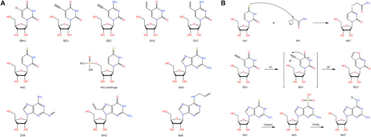
    1. Erhard F. Saliba A.-E. Lusser A. Toussaint C. Hennig T. Prusty B. K. Kirschenbaum D. Abadie K. Miska E. A. Friedel C. C. Amit I. Micura R. Dölken L. Nat. Rev. Methods Primers. 2022;2:77. doi: 10.1038/s43586-022-00157-z. - DOI
    1. Spanjaard B. Hu B. Mitic N. Olivares-Chauvet P. Janjuha S. Ninov N. Junker J. P. Nat. Biotechnol. 2018;36:469–473. doi: 10.1038/nbt.4124. - DOI - PMC - PubMed
    1. Cao J. Zhou W. Steemers F. Trapnell C. Shendure J. Nat. Biotechnol. 2020;38:980–988. doi: 10.1038/s41587-020-0480-9. - DOI - PMC - PubMed

LinkOut - more resources