[Preprint]. 2024 Nov 6:2024.11.04.621940.
doi: 10.1101/2024.11.04.621940.

Characterization of A Bronchoscopically Induced Transgenic Lung Cancer Pig Model for Human Translatability

Affiliations

Characterization of A Bronchoscopically Induced Transgenic Lung Cancer Pig Model for Human Translatability

Kirtan Joshi et al. bioRxiv. .

Abstract

Background: There remains a need for animal models with human translatability in lung cancer (LC) research. Findings in pigs have high impact on humans due to similar anatomy and physiology. We present the characterization of a bronchoscopically-induced LC model in Oncopigs carrying inducible KRASG12D and TP53R167H mutations.

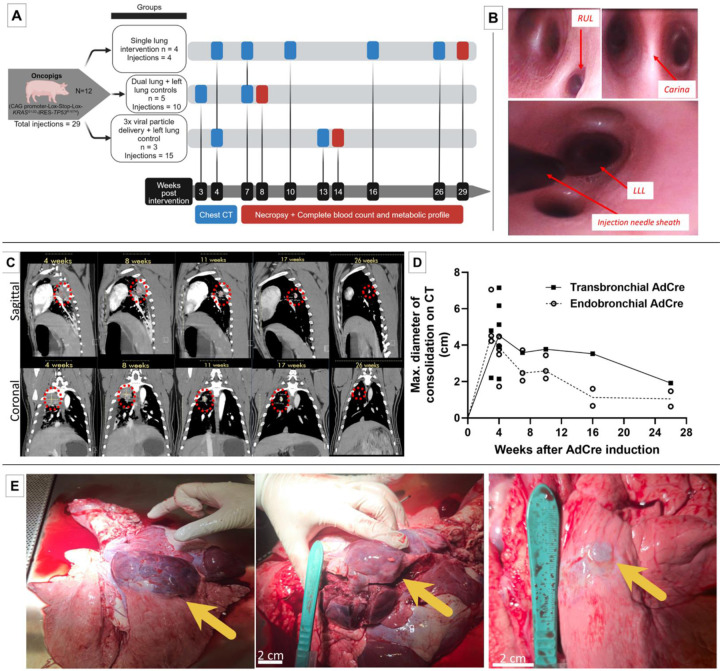
Methods: Twelve Oncopigs underwent 29 injections via flexible bronchoscopy. Eighteen Adenovirus-Cre recombinase gene (AdCre) inductions were performed endobronchially (n=6) and transbronchially with a needle (n=12). Eleven control injections were performed without AdCre. Oncopigs underwent serial contrast-enhanced chest CT with clinical follow-up for 29 weeks. Following autopsy, lung and organ tissues underwent histopathology, immunohistochemistry, and RNA-sequencing with comparative analysis with The Cancer Genome Atlas (TCGA) human LC data.

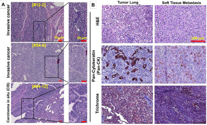
Results: All 18 sites of AdCre injections had lung consolidations on CT imaging. Transbronchial injections led to histopathologic invasive cancer and/or carcinoma in situ (CIS) in 11/12 (91.7%), and invasive cancer (excluding CIS) in 8/12 (66.6%). Endobronchial inductions led to invasive cancer in 3/6 (50%). A soft tissue metastasis was observed in one Oncopig. Immunohistochemistry confirmed expression of Pan-CK+/epithelial cancer cells, with macrophages and T cells infiltration in the tumor microenvironment. Transcriptome comparison showed 54.3% overlap with human LC (TCGA), in contrast to 29.88% overlap of KRAS-mutant mouse LC with human LC.

Conclusions: The transgenic and immunocompetent Oncopig model has a high rate of LC following bronchoscopic transbronchial induction. Overlap of the Oncopig LC transcriptome with human LC transcriptome was noted. This pig model is expected to have high clinical translatability to the human LC patient.

Keywords: Oncopig; large animal cancer model; pig lung cancer model; transgenic cancer model; translational cancer research.


Figures

Figure 1:
Figure 1:
A: Study design: Bronchoscopic inductions (total n=29) were performed in twelve Oncopigs separated into three groups. Clinical surveillance and follow-up imaging were performed with contrast-enhanced chest CT at the given time points. Blood analyses and necropsies were performed a three different time points post-induction. B: Flexible bronchoscopy images from Oncopig airways. Left upper panel: Right upper lobe (RUL) bronchus that is located proximally to the main carina. Right upper panel: Close-up view of the main carina with bifurcation into right and left main stem bronchus. Lower panel: Bronchoscopic injection needle sheath visible in the left lower lung lobe (LLL). All interventions in pigs were performed with human-grade bronchoscopy equipment. C: High-resolution, contrast-enhanced chest CT scans following bronchoscopic AdCre induction in Oncopigs. Sagittal and coronal images showing consolidation in the lung at the AdCre injection sites at various timepoints. Regression of the consolidations were observed over subsequent imaging, with persistence of solid lung nodules. Contralateral lung lobe injections with adjuvant agents (IL-8, polybrene) without AdCre did not show any consolidations on CT imaging. D: Curves showing radiographic consolidation on CT imaging determined by maximum consolidation diameter (cm) measurements according to the AdCre injection techniques (endo- versus transbronchial). (Connecting line: medians). E: Autopsy images from Oncopigs that had undergone LC induction with bronchoscopic injection of AdCre. Left panel: Right upper lobe lung (RUL) mass; Middle panel: Same dissected tumor; Right panel: Right upper lobe (RUL) nodule. (Arrows indicate tumors).
Figure 2:
Figure 2:
A: Hematoxylin and eosin (H&E) staining of three different Oncopig lung tumors showing invasive cancer cells (upper and middle panels) and carcinoma in situ (CIS) (lower panel). (Scale bars, 50 μm). B: Histopathology and immunostaining of matched Oncopig LC (left panels) and soft tissue metastasis (right panels) stained with H&E (top row), Pan-Cytokeratin (Pan-CK) for epithelial cells (middle row), and Masson’s Trichrome for visualization of connective tissue/stroma (lower row). (Scale bars, 150 μm).
Figure 3:
Figure 3:
A: Immune cell infiltration in the Oncopig LC tumor microenvironment at three different autopsy dates at 8, 14 and 29 weeks. Immunohistochemistry staining for IBA-1 (macrophages) and CD3 (T cells) identified immune cells in LCs and adjacent normal lungs, while the extent of infiltration subsequently decreased. Pan-CK staining is also shown. (Scale bars, 100 μm.). B: T cell and macrophage infiltration around invasive cancer cells in the tumor microenvironment of an Oncopig LC at higher magnification. Histopathology (H&E) staining and immunostaining for identification of Pan-CK+ epithelial cells, in addition to IBA-1+ macrophages and CD3+ T cells in the tumor microenvironment are shown. (Scale bars, 20 μm).
Figure 4.
Figure 4.
Oncopig-derived LC and soft tissue metastasis (matched) showing decreased expression of E-cadherin (2nd row) in the metastasis. Nuclear expression of proliferation marker Ki-67 (3rd row) and epithelial-to-mesenchymal transition (EMT) marker vimentin (4th row) was observed in both primary LC and the metastasis. Pan-CK expression is also shown (upper row). (Scale bars, 100 μm).
Figure 5.
Figure 5.. Whole transcriptome analysis of Oncopig lung tumors and healthy pig lung in comparison with human LC TCGA data.
A: Principal component analysis of Oncopig-derived LC and normal Oncopig lung showing similarities and variabilities between normal and tumor tissues (n=2, respectively). B: Differential gene expression analysis between LC tumor versus normal pig lung tissues. C: Gene ontology and pathway enrichment analysis of top 50 differentially expressed genes of Oncopig LCs. D: Orthologous gene expression comparison with human LC patients was performed using The Cancer Genome Atlas (TCGA) data [total N=40 non-small cell lung cancer (NSCLC) patients’ transcriptomic data [N=10 per stage I-IV, respectively)], demonstrating 54.3% of the Oncopig LC transcripts overlapping with human LC transcripts, in contrast to less (29.88%) overlap with mouse KRAS-mutant LC.

References

    1. Tanaka R. et al. A mouse model of lung cancer induced via intranasal injection for anticancer drug screening and evaluation of pathology. FEBS Open Bio 13, 51–59 (2023). - PMC - PubMed
    1. Mak I.W., Evaniew N. & Ghert M. Lost in translation: animal models and clinical trials in cancer treatment. Am J Transl Res 6, 114–118 (2014). - PMC - PubMed
    1. Holliday R. Neoplastic transformation: the contrasting stability of human and mouse cells. Cancer Surv 28, 103–115 (1996). - PubMed
    1. Kaiser J. The cancer test. Science 348, 1411–1413 (2015). - PubMed
    1. Pan Z. et al. Pig genome functional annotation enhances the biological interpretation of complex traits and human disease. Nat Commun 12, 5848 (2021). - PMC - PubMed

Publication types