

Progress of fracture mapping technology based on CT three-dimensional reconstruction
- PMID: 39569162
- PMCID: PMC11576209
- DOI: 10.3389/fbioe.2024.1471470
Progress of fracture mapping technology based on CT three-dimensional reconstruction
Abstract
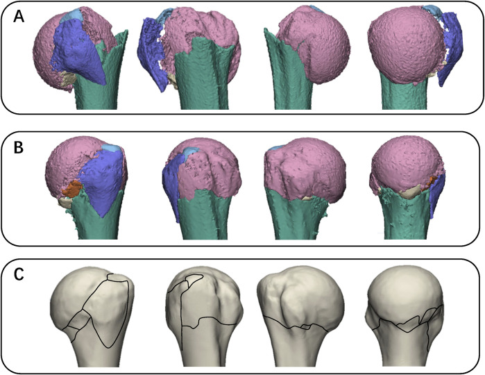
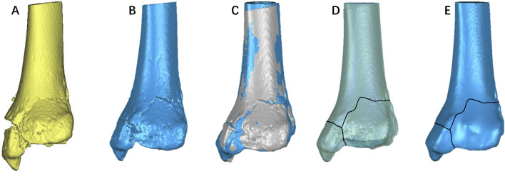
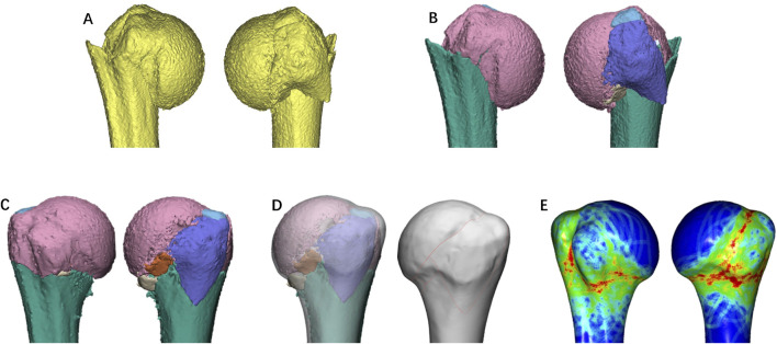
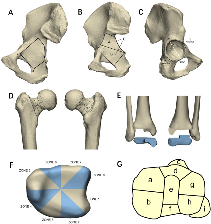
Fracture Mapping is a new technology developed in recent years. This technology visually representing the morphology of fractures by overlaying fracture lines from multiple fracture models onto a standard model through three-dimensional reconstruction. Fracture mapping has been widely used in acetabular fracture, proximal humerus fractures, Pilon fracture, tibial plateau fractures, and so on. This technology provides a new research method for the diagnosis, classification, treatment selection, internal fixation design, and statistical analysis of common fracture sites. In addition, the fracture map can also provide a theoretical basis for the establishment of a biomechanical standardized fracture model. Herein, we reviewed various methods and the most advanced techniques for fracture mapping, and to discuss the issues existing in fracture mapping techniques, which will help in designing future studies that are closer to the ideal. Moreover, we outlined the fracture morphology features of fractures in various parts of the body, and discuss the implications of these fracture mapping studies for fracture treatment, thereby providing reference for research and clinical decision-making on bone and joint injuries to improve patient prognosis.
Keywords: classification; fracture mapping; fractures; heat map; morphology; three-dimensional.
Copyright © 2024 Liu, Zhang, Qu and Piao.
Conflict of interest statement
The authors declare that the research was conducted in the absence of any commercial or financial relationships that could be construed as a potential conflict of interest.
Figures
Similar articles
-
Three-dimensional mapping of distal humerus fracture.J Orthop Surg Res. 2021 Sep 3;16(1):545. doi: 10.1186/s13018-021-02691-0. J Orthop Surg Res. 2021. PMID: 34479569 Free PMC article.
-
Tibial plateau fractures: three dimensional fracture mapping and morphologic measurements.Int Orthop. 2022 Sep;46(9):2153-2163. doi: 10.1007/s00264-022-05434-w. Epub 2022 May 17. Int Orthop. 2022. PMID: 35579696 Free PMC article.
-
[Research progress on biomechanics for internal fixation in tibial plateau fracture].Zhongguo Xiu Fu Chong Jian Wai Ke Za Zhi. 2024 Jan 15;38(1):113-118. doi: 10.7507/1002-1892.202309077. Zhongguo Xiu Fu Chong Jian Wai Ke Za Zhi. 2024. PMID: 38225850 Free PMC article. Review. Chinese.
-
Tibial Plateau Fracture Characteristics: Computed Tomography Mapping of Lateral, Medial, and Bicondylar Fractures.J Bone Joint Surg Am. 2015 Sep 16;97(18):1512-20. doi: 10.2106/JBJS.N.00866. J Bone Joint Surg Am. 2015. PMID: 26378267
-
High-energy pilon fractures management: State of the art.EFORT Open Rev. 2017 Mar 13;1(10):354-361. doi: 10.1302/2058-5241.1.000016. eCollection 2016 Oct. EFORT Open Rev. 2017. PMID: 28461913 Free PMC article. Review.
Cited by
-
Mapping of Patellar Fracture Patterns: A Multicenter Study of 237 Patients.J Clin Med. 2025 Feb 17;14(4):1335. doi: 10.3390/jcm14041335. J Clin Med. 2025. PMID: 40004864 Free PMC article.
References
-
- Adelaar R. S. (1997). Complex fractures of the talus. Instr. Course Lect. 46, 323–338. - PubMed
Publication types
LinkOut - more resources
Full Text Sources