Review
. 2024 Nov 6:12:1441430.
doi: 10.3389/fbioe.2024.1441430. eCollection 2024.

Surface-modified CMOS biosensors

Affiliations
Review

Surface-modified CMOS biosensors

Fahimeh Dehghandehnavi et al. Front Bioeng Biotechnol. .

Abstract

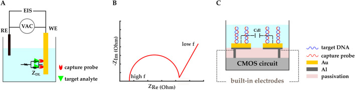
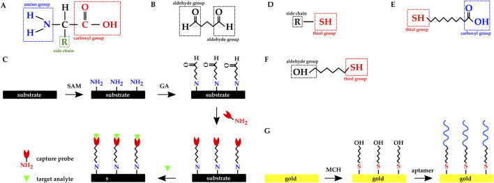
Biosensors translate biological events into electronic signals that quantify biological processes. They are increasingly used in in vitro diagnostics applications that leverage their ability to process small sample volumes. One recent trend has been to integrate biosensors with complementary metal-oxide-semiconductor (CMOS) chips to provide enhanced miniaturization, parallel sensing, and low power consumption at a low cost. CMOS-enabled biosensors are used in monitoring DNA hybridization, enzymatic reactions, and cell proliferation, to name a few applications. This paper explores the materials and processes used in emerging CMOS biosensors. We discuss subtractive and additive processes for creating electrodes for electrochemical sensing applications. We discuss functionalization techniques for creating bioelectronic interfaces that allow molecular events to be transduced into the electrical domain using a plurality of modalities that are readily provided by CMOS chips. Example modalities featured are optical sensing, electrochemical detection, electrical detection, magnetic sensing, and mechanical sensing.

Keywords: biosensor; complementary metal-oxide-semiconductor (CMOS); immobilization; lab-on-a-chip (LOC); post-CMOS process; transduction.


Conflict of interest statement

The authors declare that the research was conducted in the absence of any commercial or financial relationships that could be construed as a potential conflict of interest.

Figures

FIGURE 1
FIGURE 1
Schematic representation of biosensors’ working principle.
FIGURE 2
FIGURE 2
Schematic representation of functionalization mechanisms. (A) Physical adsorption. (B) Streptavidin-biotin complex. (C) Covalent immobilization.
FIGURE 3
FIGURE 3
Schematic representation and circuit equivalent of (A, C) two-electrode and (B, D) three-electrode electrochemical biosensing. (E) CMOS electrochemical biosensor. Rsn is the solution equivalent resistance between the RE and the BRE. ZCE is the equivalent impedance between the CE and the RE. Isn is the current going through the solution. Zbio represents the BRE equivalent impedance.
FIGURE 4
FIGURE 4
Schematic representation of (A) amperometric/voltammetric electrochemical biosensor. The TIA is a transimpedance amplifier. (B) An example of cyclic voltammetry I-V plot. (C) Amperometric CMOS biosensor developed by (Manickam et al., 2019a; b). Electrodes were realized by covering the exposed top metal layer with a-C. A 10 nm Ti layer was used as the adhesion layer. MB-labeled stem-loop DNA hairpin probes were covalently immobilized on the a-C electrodes.
FIGURE 5
FIGURE 5
(A) Schematic representation of a potentiometric electrochemical biosensor. Δ V is an open-circuit voltage between RE and WE.(B) An ISE-based potentiometric CMOS biosensor developed by (Doi et al., 2022).
FIGURE 6
FIGURE 6
(A) Schematic representation of an impedimetric electrochemical biosensor. ZDL and EIS are double-layer impedance and electrochemical impedance spectroscopy, respectively. (B) An example Nyquist plot. (C) A CMOS capacitive biosensor designed by (Kuo et al., 2020). Electrodes were initially made with the top metal layer and they were covered by Au in post-processing. Thiolated microRNA–195 probes were immobilized on the gold IDE. Cdl represents the double layer capacitance.
FIGURE 7
FIGURE 7
(A) Schematic representation of FET-based CMOS electrochemical biosensor (B) A CMOS ISFET-based biosensor developed by (Saengdee et al., 2021). Anti-HSA was covalently bonded on the Si3Ni4 passivation layer through the APTES-GA method to function as the sensing membrane. An Ag/AgCl electrode was used as the RE. The Si3Ni4 -ISFET current drain was kept constant and the gate potential was monitored.
FIGURE 8
FIGURE 8
(A) EG-FET biosensor introduced by (Sheibani et al., 2021). The extended gate was a single-layer graphene on a platinum electrode. and it was connected to the MOSFET’s metal gate with metal vias. The extended gate and the Ag/AgCl reference electrode were exposed to the analyte solution, while the MOSFET transducer was isolated from the liquid under testing. Cortisol aptamers were immobilized on the graphene. (B) Schematic representation of a Si NW FET. The Si NW replaces the gate and the doped channel in regular MOSFETs, and the NW surface functions as the sensing membrane. Molecular probes are commonly immobilized on the NW’s surface.
FIGURE 9
FIGURE 9
CMOS optical biosensing binding mechanisms:(A) direct. Capture probes are immobilized on the CMOS chip’s surface and target molecules are either inherently fluorescent or pre-labeled with a fluorescent group. (B) sandwich. Capture probes are immobilized on the sensor’s active region and fluorescent tracer molecules are added to the analytic solution. Target molecules bind to the capture probe and the fluorescent tracer molecule at the same time. Free fluorescent tracer molecules are physically removed from the solution or optically excluded from the sensing region. The remaining fluorescent signal is an indicator of the target biomolecule. (C) competitive. Capture probes are immobilized on the chip’s surface and pre-labeled target molecules are added to the sample solution while the target molecules in the analytic solution are not labeled. The added labeled molecules compete with the unlabeled molecules over the available binding sites. The fluorescent signal decreases as the concentration of the target molecules in the sample under test increases.
FIGURE 10
FIGURE 10
Schematic representation of the CMOS electrical biosensor developed by (Hall et al., 2022); (A) a peptide molecular wire connecting two Ru nanoelectrodes, (B) a series of current pulses indicating binding events happening. (C) CMOS magnetic biosensor developed by (Costa et al., 2017); SV sensors were post-fabricated on top of a CMOS IC front-end and the sensors were covered by an AlN passivation layer.
FIGURE 11
FIGURE 11
CMOS microcantilever-based biosensors: (A) the cantilever’s surface was oxidized and AFP capture antibodies were covalently immobilized on it via APTES-GA crosslinking. AuNPs were functionalized with detection antibodies (Zhao et al., 2021); (B) Vp capture antibodies were immobilized on the Au electrodes fabricated on the CMOS microcantilever (Wang et al., 2021). (C) CMOS multimodal biosensor developed by (Al-Rawhani et al., 2020).
FIGURE 12
FIGURE 12
Chemical structure of (A) Amino acid. (B) Glutaraldehyde molecule. (C) Overview of the dielectric substrate preparation with SAM and GA molecules: amine functionalization, activation with glutaraldehyde, probe immobilization, and immunoassay. Chemical structure of (D) a molecule with thiol group. (E) 11-mercaptoundecanoic acid. (F) 6 mercapto 1 hexanol. (G) Overview of the gold electrode’s functionalization with MCH SAM and thiolated aptamer.

References

    1. Adem S., Jain S., Sveiven M., Zhou X., O’Donoghue A. J., Hall D. A. (2020). Giant magnetoresistive biosensors for real-time quantitative detection of protease activity. Sci. Rep. 10, 7941. 10.1038/s41598-020-62910-2 - DOI - PMC - PubMed
    1. Akçapınar R., Garipcan B., Goodarzi V., Uzun L. (2021). Designing of various biosensor devices for determination of apoptosis: a comprehensive review. Biochem. Biophysical Res. Commun. 578, 42–62. 10.1016/j.bbrc.2021.08.089 - DOI - PubMed
    1. Al-Rawhani M. A., Mitra S., Barrett M. P., Cochran S., Cumming D. R., Hu C., et al. (2020). Multimodal integrated sensor platform for rapid biomarker detection. IEEE Trans. Biomed. Eng. 67, 614–623. 10.1109/TBME.2019.2919192 - DOI - PubMed
    1. Ambhorkar P., Wang Z., Ko H., Lee S., Koo K. I., Kim K., et al. (2018). Nanowire-based biosensors: from growth to applications. Micromachines 9, 679. 10.3390/mi9120679 - DOI - PMC - PubMed
    1. Anand U., Chandel A. K. S., Oleksak P., Mishra A., Krejcar O., Raval I. H., et al. (2022). Recent advances in the potential applications of luminescence-based, SPR-based, and carbon-based biosensors. Appl. Microbiol. Biotechnol. 106, 2827–2853. 10.1007/s00253-022-11901-6 - DOI - PMC - PubMed

LinkOut - more resources