. 2024 Nov 6:26:1048-1057.
doi: 10.1016/j.reth.2024.10.010. eCollection 2024 Jun.

Platelet-rich plasma-derived extracellular vesicles improve liver cirrhosis in mice

Affiliations

Platelet-rich plasma-derived extracellular vesicles improve liver cirrhosis in mice

Yuichirou Maeda et al. Regen Ther. .

Abstract

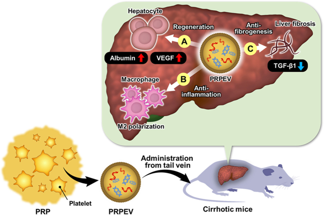
Introduction: Cirrhosis remains a significant clinical challenge due to its poor prognosis and limited treatment options, creating a high unmet medical need for the development of novel therapies. In this study, we analyzed the effects of a novel approach to treat cirrhosis using platelet-rich plasma-derived extracellular vesicles (PRPEV) in mice.

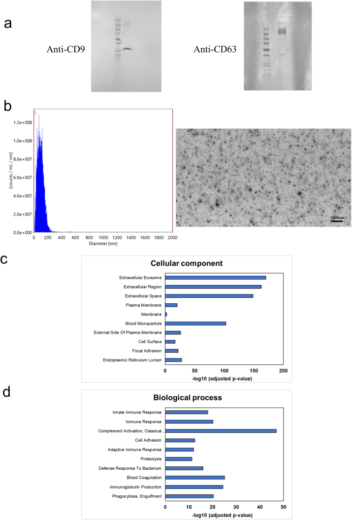
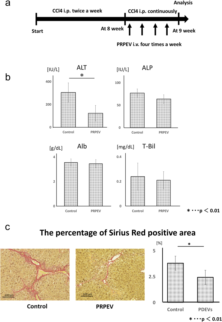
Methods: PRPEV were collected from platelet-rich plasma using ultrafiltration, and their proteomes were analyzed. The carbon tetrachloride (CCl4)-induced cirrhosis model of mice was used to evaluate the effect of PRPEV administration and compared with the control group (n = 8). In vitro and in vivo mechanistic analyses of PRPEV administration were confirmed using real time-PCR and immunostaining.

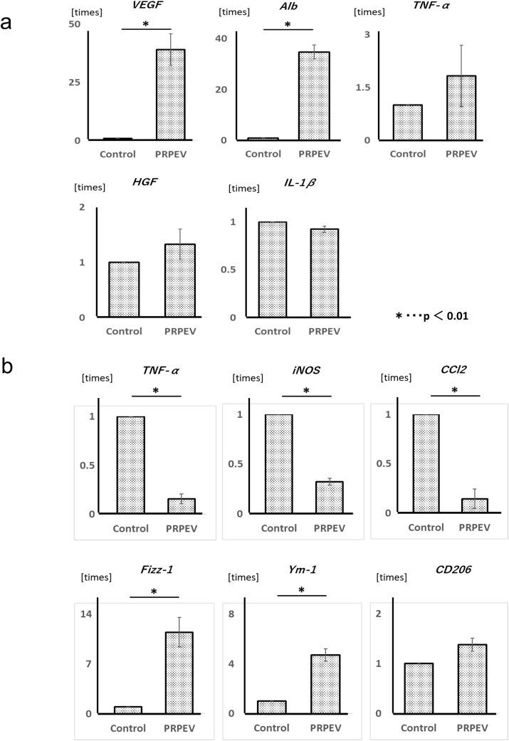
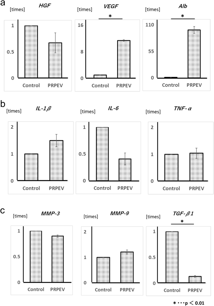
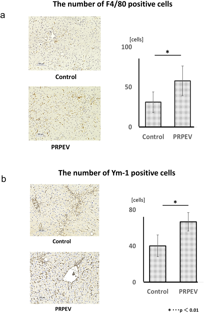
Results: Gene ontology analysis based on the proteome revealed that PRPEV contain many factors associated with EV and immune responses. In vitro, PRPEV polarize macrophages into an anti-inflammatory phenotype. Following PRPEV administration, there was a decrease in serum alanine aminotransferase levels and reduction in liver fibrosis, while mRNA levels of regenerative factors were upregulated and transforming growth factor β-1 was downregulated. Furthermore, the number of anti-inflammatory macrophages in the liver increased.

Conclusions: PRPEV may contribute to hepatocyte proliferation, anti-inflammation, and anti-fibrogenesis in the liver. This novel concept paves the way for cirrhosis treatment.

Keywords: Liver cirrhosis; Platelet; Platelet-rich plasma; Platelet-rich plasma-derived extracellular vesicles.


Conflict of interest statement

None.

Figures

Image 1
Graphical abstract
Fig. 1
Fig. 1
Identification and characteristics of platelet-rich plasma-derived extracellular vesicles (PRPEV). (a) Western blot analysis of CD 9 and CD 63 of PRPEV; n = 3 for each experiment. (b) Particle size distribution of PRPEV; n = 3 for each experiment. (c–d) Gene ontology analysis based on the proteome of PRPEV. The top 10 enriched terms in the Cellular Component and Biological Process are represented.
Fig. 2
Fig. 2
In vitro analysis using hepatocytes and macrophages. (a) Changes in the mRNA expression of vascular endothelial growth factor (VEGF), albumin (Alb), Tumor necrosis factor (TNF) -α, hepatocyte growth factor (HGF), and interleukin (IL) -1β in the hepatocytes (AML12) after addition of platelet-rich plasma-derived extracellular vesicles (PRPEV); n = 6 for each group. Data are presented as ratio to the standards as the mean ± SD, p < 0.01 (VEGF, PRPEV compared to Control), p < 0.01 (Alb, PRPEV compared to Control). (b) Changes in the mRNA expression of TNF-α, induced nitric oxide synthase (iNOS), chemokine (C–C motif) ligand 2 (CCl2), Fizz-1, Ym-1, and CD206 in the macrophages after addition of PRPEV; n = 6 for each group. Data are presented ratio to the standards as the mean ± SD, p < 0.01 (TNF-α, PRPEV compared to Control), p < 0.01 (iNOS, PRPEV compared to Control), p < 0.01 (CCl2, PRPEV compared to Control), p < 0.01 (Fizz-1, PRPEV compared to Control), p < 0.01 (Ym-1, PRPEV compared to Control).
Fig. 3
Fig. 3
Therapeutic effect of platelet-rich plasma-derived extracellular vesicles (PRPEV) on cirrhosis in mice. (a) Schematic representation of the timeline from PRPEV administration to analysis of cirrhotic model mice. (b) Serum levels of alanine aminotransferase (ALT), alkaline phosphatase (ALP), albumin (Alb), and total bilirubin (T-Bil) after PRPEV administration. Data are presented as the mean ± SD, n = 8 in each group, p < 0.01 (ALT, PRPEV compared to Control). (c) Immunostaining with Sirius Red in the liver showing the degree of liver fibrosis in mice from Control and PRPEV group; n = 8 for each group. Data are presented as the mean ± SD, p < 0.01 (PRPEV compared to Control).
Fig. 4
Fig. 4
Changes in the mRNA expression after platelet-rich plasma-derived extracellular vesicles (PRPEV) administration in the whole liver. Data are presented ratio to the standards as the mean ± SD, n = 8 for each group. (a) Changes in the mRNA expression related to liver regeneration (hepatocyte growth factor [HGF], vascular endothelial growth factor [VEGF], and albumin [Alb]). p < 0.01 (VEGF, PRPEV compared to Control). (b) Changes in the mRNA expression related to inflammation (interleukin-1β [IL-1β], interleukin-6 [IL-6], and tumor necrosis factor-α [TNF-α]). (c) Changes in the mRNA expression related to liver fibrosis (matrix metalloplotainase-3 [MMP-3], matrix metalloplotainase-9 [MMP-9], and transforming growth factor-β1 [TGF-β1]). p < 0.01 (TGF-β1, PRPEV compared to Control).
Fig. 5
Fig. 5
Analysis of macrophages in the liver after platelet-rich plasma-derived extracellular vesicles (PRPEV) administration. (a) Immunostaining for F4/80 in the liver; n = 8 for each group. The number of F4/80 positive cells in the liver was counted, and data are presented as the mean ± SD. p < 0.01 (PRPEV compared to Control). (b) Immunostaining for Ym-1 in the liver; n = 8 for each group. The number of Ym-1 positive cells in the liver was counted, and data are presented as the mean ± SD. p < 0.01 (PRPEV compared to Control).

References

    1. Asrani S.K., Devarbhavi H., Eaton J., Kamath P.S. Burden of liver diseases in the world. J Hepatol. 2019;70:151–171. - PubMed
    1. Zarrinpar A., Busuttil R.W. Liver transplantation: past, present and future. Nat Rev Gastroenterol Hepatol. 2013;10:434–440. - PubMed
    1. Terai S., Tsuchiya A., Watanabe Y., Takeuchi S. Transition of clinical and basic studies on liver cirrhosis treatment using cells to seek the best treatment. Inflamm Regen. 2021;41:27. - PMC - PubMed
    1. Lucey M.R., Furuya K.N., Foley D.P. Liver transplantation. N Engl J Med. 2023;389:1888–1900. - PubMed
    1. Underwood P.W., Cron D.C., Terjimanian M.N., Wang S.C., Englesbe M.J., Waits S.A. Sarcopenia and failure to rescue following liver transplantation. Clin Transplant. 2015;29:1076–1080. - PubMed

LinkOut - more resources