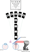
Accommodative behaviour and retinal defocus in highly myopic eyes fitted with a dual focus myopia control contact lens
- PMID: 39569764
- PMCID: PMC11629841
- DOI: 10.1111/opo.13420
Accommodative behaviour and retinal defocus in highly myopic eyes fitted with a dual focus myopia control contact lens
Abstract
Purpose: To evaluate the myopic and hyperopic defocus delivered to the retina by a dual focus (DF) myopia control contact lens when myopia exceeds 6.00 D.
Methods: Individuals with high myopia were fitted bilaterally with high-powered DF lenses containing power profiles matching a Coopervision MiSight 1 day contact lens (omafilcon A) and a Coopervision Proclear 1 day single vision (SV) lens. Wavefront measurements along the primary line of sight and across the central ±20° of the horizontal retina were acquired using a pyramidal aberrometer, while subjects accommodated to high-contrast letter stimuli (6/12 equivalent) at six target vergences (-0.25 and -1.00 to -5.00 D). Linear mixed-effects regression models explored the relationship between the spherical equivalent refractive error (SERE) and induced defocus.
Results: Thirteen teenagers and young adults (ages 13-32 years, mean [standard deviation, SD] age = 22.8 [4.9] years) with high myopia (SERE -6.50 to -9.25 D) were tested. The treatment optic zone of the DF lens shifted retinal defocus by the expected -2.00 D, with a mean (SD) difference (DF-SV) of -2.21 (0.18) D for the inner treatment ring. Inclusion of the treatment optic had no significant impact on accommodative accuracy (p = 0.51). Accommodative lags were larger at the nearer viewing distances, with lag increasing by approximately 0.30 D for every additional dioptre of SERE. Measured retinal defocus within the annular treatment zone was approximately -2.00 D at the foveal centre, 10° nasal and temporal and 20° nasal and reduced to -1.90 (0.57) D at 20° temporal.
Conclusions: Relative to eyes with lower levels of myopia, the increased accommodative lags and more prolate retinas of highly myopic eyes reduced the myopic retinal defocus from the DF myopia control lens, while the treatment optical zones generated the combined effect of reducing hyperopic and introducing myopic retinal defocus relative to an SV correction.
Keywords: accommodative behaviour; contact lenses; dual focus contact lenses; high myopia; myopia control; retinal defocus.
© 2024 The Author(s). Ophthalmic and Physiological Optics published by John Wiley & Sons Ltd on behalf of College of Optometrists.
Conflict of interest statement
DM: none; JG: none; MR: consultant: CooperVision Inc., SightGlass Vision; NM: none; PC: employee, CooperVision Inc.; AB: employee, CooperVision Inc. PK: research: Alcon, CooperVision Inc., EssilorLuxottica, Hoya, Johnson and Johnson Vision, SightGlass Vision; PK: consultant EssilorLuxottica, SightGlass Vision.
Figures


Similar articles
-
Accommodative behaviour and retinal defocus of children during prolonged viewing of electronic devices while wearing dual-focus myopia control soft contact lenses.Ophthalmic Physiol Opt. 2025 Sep;45(6):1456-1467. doi: 10.1111/opo.13533. Epub 2025 May 28. Ophthalmic Physiol Opt. 2025. PMID: 40437791 Free PMC article.
-
Retinal defocus in myopes wearing dual-focus zonal contact lenses.Ophthalmic Physiol Opt. 2022 Jan;42(1):8-18. doi: 10.1111/opo.12903. Epub 2021 Oct 23. Ophthalmic Physiol Opt. 2022. PMID: 34687238 Free PMC article.
-
Myopia Control Dose Delivered to Treated Eyes by a Dual-focus Myopia-control Contact Lens.Optom Vis Sci. 2023 Jun 1;100(6):376-387. doi: 10.1097/OPX.0000000000002021. Epub 2023 Apr 24. Optom Vis Sci. 2023. PMID: 37097975 Free PMC article.
-
Astigmatic Peripheral Defocus with Different Contact Lenses: Review and Meta-Analysis.Curr Eye Res. 2016 Aug;41(8):1005-1015. doi: 10.3109/02713683.2015.1116585. Epub 2016 Feb 2. Curr Eye Res. 2016. PMID: 26835871 Review.
-
Lenses and Spectacles to Prevent Myopia Worsening in Children [Internet].Ottawa (ON): Canadian Agency for Drugs and Technologies in Health; 2021 Apr. Ottawa (ON): Canadian Agency for Drugs and Technologies in Health; 2021 Apr. PMID: 34255449 Free Books & Documents. Review.
Cited by
-
Accommodative behaviour and retinal defocus of children during prolonged viewing of electronic devices while wearing dual-focus myopia control soft contact lenses.Ophthalmic Physiol Opt. 2025 Sep;45(6):1456-1467. doi: 10.1111/opo.13533. Epub 2025 May 28. Ophthalmic Physiol Opt. 2025. PMID: 40437791 Free PMC article.
References
-
- Jung SK, Lee JH, Kakizaki H, Jee D. Prevalence of myopia and its association with body stature and educational level in 19‐year‐old male conscripts in Seoul, South Korea. Invest Ophthalmol Vis Sci. 2012;53:5579–5583. - PubMed
-
- Sun J, Zhou J, Zhao P, Lian J, Zhu H, Zhou Y, et al. High prevalence of myopia and high myopia in 5060 Chinese university students in Shanghai. Invest Ophthalmol Vis Sci. 2012;53:7504–7509. - PubMed
-
- Holden BA, Fricke TR, Wilson DA, Jong M, Naidoo KS, Sankaridurg P, et al. Global prevalence of myopia and high myopia and temporal trends from 2000 through 2050. Ophthalmology. 2016;123:1036–1042. - PubMed
-
- Willis JR, Vitale S, Morse L, Parke DW 2nd, Rich WL, Lum F, et al. The prevalence of myopic choroidal neovascularization in the United States: analysis of the IRIS(®) data registry and NHANES. Ophthalmology. 2016;123:1771–1782. - PubMed
-
- Lin LL, Shih YF, Hsiao CK, Chen CJ. Prevalence of myopia in Taiwanese schoolchildren: 1983 to 2000. Ann Acad Med Singapore. 2004;33:27–33. - PubMed
MeSH terms
Grants and funding
LinkOut - more resources
Full Text Sources
Miscellaneous

