. 2025 May 15;27(4):932-947.
doi: 10.1093/neuonc/noae248.

G-quadruplex stabilizer CX-5461 effectively combines with radiotherapy to target α-thalassemia/mental retardation X-linked-deficient malignant glioma

Affiliations

G-quadruplex stabilizer CX-5461 effectively combines with radiotherapy to target α-thalassemia/mental retardation X-linked-deficient malignant glioma

Sharvari Dharmaiah et al. Neuro Oncol. .

Abstract

Background: Inactivation of α-thalassemia/mental retardation X-linked (ATRX) represents a defining molecular feature in large subsets of malignant glioma. ATRX deficiency gives rise to abnormal G-quadruplex (G4) DNA secondary structures, enhancing replication stress and genomic instability. Building on earlier work, we evaluated the extent to which pharmacological G4 stabilization selectively enhances DNA damage and cell death in ATRX-deficient preclinical glioma models.

Methods: Using the G4 stabilizer CX-5461, we treated patient-derived glioma stem cells (GSCs) in vitro and GSC flank and intracranial murine xenografts in vivo to evaluate efficacy as both a single agent and in combination with ionizing radiation (IR), the latter a central element of current treatment standards.

Results: CX-5461 promoted dose-sensitive lethality in ATRX-deficient GSCs relative to ATRX-intact controls. Mechanistic studies revealed that CX-5461 disrupted histone variant H3.3 deposition, enhanced replication stress and DNA damage, activated p53-independent apoptosis, and induced G2/M arrest to a greater extent in ATRX-deficient GSCs than in ATRX-intact counterparts. These data were corroborated in vivo, where CX-5461/IR treatment profoundly delayed tumor growth and prolonged survival in mice bearing ATRX-deficient flank xenografts. Histopathological analyses revealed decreased proliferation, increased apoptosis, and significant G4 induction, replication stress, and DNA damage in CX-5461-treated tumors, both alone and in combination with IR. Finally, despite suboptimal blood-brain-barrier penetration, systemic CX-5461 treatment induced tangible pharmacodynamic effects in ATRX-deficient intracranial GSC models.

Conclusions: In totality, our work substantively demonstrates efficacy and defines mechanisms of action for G4 stabilization as a novel therapeutic strategy targeting ATRX-deficient malignant glioma, laying the groundwork for clinical translation.

Keywords: ATRX; CX-5461; G-quadruplex; glioma; radiation.


Conflict of interest statement

None declared.

Figures
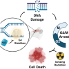
Graphical Abstract
Graphical Abstract
Figure 1.
Figure 1.
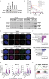
CX-5461 increases G4s, replication stress, and DNA damage in primary patient-derived ATRX-deficient glioma stem cells (GSCs). (A) Western blot validation of ATRX status in GSC lines (β-actin loading control). (B) Cell viability curves for the indicated GSCs showing increased CX-5461 sensitivity in ATRX-deficient variants (TS 543-sh590, TS 603-shATRX, GS 5-22, and GS 8-18). Cell viability was measured 5 days after initiation of treatment with CX-5461 using Promega® CellTiter-Glo. (C) Western blots showing induction of replication stress (ph-ATR, ph-CHK1) and DNA damage (ph-ATM, ph-CHK2) pathway markers (vinculin loading control) in ATRX-deficient (TS 543-sh590) and ATRX-intact (TS 543) GSCs upon CX-5461 treatment (1 µM) at the indicated times (h: hours). (D) Representative IF images showing foci and colocalization of G4s (BG4), RPA32, and γH2AX in ATRX-deficient (TS 543-sh590) and ATRX-intact (TS 543) GSCs treated with either vehicle or CX-5461 (1 µM; 60× magnification). (E) Quantification of IF staining for BG4, RPA32, and γH2AX nuclear foci in GSCs (N = 30 single cell images). (F) Gene Set Enrichment Analysis (GSEA) hallmark pathways positively and negatively enriched in CX-5461-treated ATRX-deficient (TS 543-sh590) GSCs. (G) GSEA plot documenting induction of DNA damage repair GSEA Hallmark pathway. Dotted line demarcates the 0.0 enrichment score. All experiments were conducted 48 hours post-CX-5461 treatment (1 µM), unless indicated otherwise. * P < .05, ** P < .01, *** P < .001, and **** P < .0001.
Figure 2.
Figure 2.
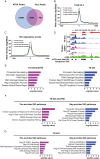
H3.3 deposition at Putative Quadruplex Sequences (PQS) is disrupted by ATRX deficiency and G4 stabilization. (A) Venn diagram depicting shared and specific peaks between H3.3 (CUT&Tag) and ATRX (CUT&RUN) (B-C) H3.3 CUT&Tag enrichment plots for all peaks (B) and peaks overlapping PQS (C) in ATRX-deficient (TS 543-sh590) and ATRX-intact (TS 543) glioma stem cells (GSCs) treated with either vehicle or CX-5461 (1 µM; 48 hours). (D) Representative H3.3 CUT&Tag traces at the MYC locus for ATRX-deficient (TS 543-sh590) and ATRX-intact (TS 543) GSCs treated with either vehicle or CX-5461 (1 µM; 48 hours). ATRX peak (MACS2), ATRX trace (CUT&RUN), and PQS (Quadparser) positioning is also shown. (E) Enrichr gene ontology (GO) plots for relevant significantly enriched pathways at sites of differential H3.3 content (P < .05) with CX-5461 treatment (1 µM; 48 hours) in ATRX-deficient (TS 543-sh590) and ATRX-intact (TS 543) GSCs. (F-G) Enrichment plots (positive and negative) for GO pathways correlating with cross-referenced gene sets showing both differential transcription and differential H3.3 content in ATRX-deficient (TS 543-sh590) and ATRX-intact (TS 543) GSCs treated with either vehicle or CX-5461 (1 µM; 48 hours).
Figure 3.
Figure 3.
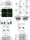
CX-5461 induces p53-independent apoptosis and G2/M cell cycle arrest in ATRX-deficient glioma stem cells (GSCs). (A) Western blots for cleaved caspase 3 (vinculin loading control) in ATRX-deficient (TS 543-sh590) and ATRX-intact (TS 543) GSCs treated with either vehicle or CX-5461 (1 µM, times indicated). (B-C) Incucyte live-cell IF imaging for cleaved caspase 3 in in ATRX-deficient (TS 543-sh590) and ATRX-intact (TS 543) GSCs treated with either vehicle or CX-5461 (1.25 µM). Representative images (B; 10× magnification) and quantified results (C; N = 8/condition) are shown. (D) Gene Set Enrichment Analysis (GSEA) plot for apoptosis GSEA Hallmark pathway in ATRX-deficient GSCs treated with CX-5461 (1 µM). The dotted line demarcates a 0.0 enrichment score. (E) Western blots for p53 and p21 (β-actin loading control) in ATRX-deficient (TS 543-sh590, GS 5-22, and GS 8-18) and ATRX-intact (TS 543) GSCs treated with either vehicle or CX-5461 (1 µM) reveal minimal p21 induction upon G4 stabilization. (F) Cell cycle flow cytometry plots for ATRX-deficient (TS 543-sh590) and ATRX-intact (TS 543) GSCs treated with either vehicle or CX-5461 (1 µM) reveal profound G2/M arrest in the ATRX-deficient context upon G4 stabilization. (G) Quantification of cell cycle flow cytometry in vehicle- and CX-5461-treated ATRX-deficient (TS 543-sh590, GS 5-22) and ATRX-intact (TS 543, TS 603) GSCs. (H) GSEA plot for the G2/M checkpoint GSEA Hallmark pathway confirming negative enrichment in ATRX-deficient TS 543-sh590 GSCs upon 1 µM CX-5461 treatment. The dotted line demarcates a 0.0 enrichment score. (I) Western blots for cyclin D1 (vinculin loading control) in ATRX-deficient (TS 543-sh590) and ATRX-intact (TS 543) GSCs treated with either vehicle or CX-5461 (1 µM; times indicated). When not stated, experiments were conducted 48 hours post-treatment with CX-5461. * P < .05, ** P < .01, *** P < .001, and **** P < .0001.
Figure 4.
Figure 4.
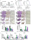
CX-5461 combines with ionizing radiation (IR) to selectively impact the in vivo growth of ATRX-deficient GSC xenografts. (A) Tumor growth curves for ATRX-deficient (TS 543-sh590) and ATRX-intact (TS 543) glioma stem cells (GSCs) flank xenografts in mice treated with either vehicle, CX-5461, IR, or combined CX-5461/IR (CX-5461: 50 mg/kg twice weekly and IR: 2 Gy × 10 fractions; treatment initiated 24 hours following GSC engraftment; N = 20/arm). (B) Kaplan–Meier survival curves for ATRX-deficient (TS 543-sh590) and ATRX-intact (TS 543) GSCs flank xenografts in mice treated with either vehicle, CX-5461, IR, or combined CX-5461/IR (CX-5461: 50 mg/kg twice weekly and IR: 2 Gy × 10 fractions; treatment initiated 24 hours following GSC engraftment; N = 15/arm). (C-F) IHC for Ki-67 and cleaved caspase 3 in harvested ATRX-deficient (TS 543-sh590) and ATRX-intact (TS 543) flank GSC xenografts treated with either vehicle, CX-5461, IR, or combined CX-5461/IR (see treatment conditions above). Representative images (C-D; H&E, 2× magnification, 20× inset; Ki67, 2× magnification, 20× inset; cleaved caspase 3, 10× magnification, 20× inset) and IHC quantification (E-F; 5 HPFs per arm; N = 5/arm) are shown. (G) Integrated density quantification of multiplexed IF for BG4, RPA32, and γH2AX in harvested ATRX-deficient (TS 543-sh590) and ATRX-intact (TS 543) flank GSC xenografts treated with either vehicle, CX-5461, IR, or combined CX-5461/IR (see treatment conditions above; N = 5/arm). Where applicable, error bars indicate SEM; P values were calculated by unpaired, parametric t-test. * P < .05, ** P < .01, *** P < .001, and **** P < .0001.
Figure 5.
Figure 5.
CX-5461 exhibits limited CNS penetration and significant pharmacodynamic effects in ATRX-deficient intracranial xenografts. (A) PK analysis depicting the plasma and brain bioavailability of CX-5461 across 24 hours, following single i.v. or p.o. administrations (i.v.: 0.3 mg/kg, p.o. plasma: 10 mg/kg, p.o. 10 mg/kg; N = 12 for i.v. and N = 30 for p.o.). (B-C) IHC for γH2AX in harvested ATRX-deficient (TS 543-sh590) intracranial GSC xenografts from mice treated with either vehicle or CX-5461 (p.o. and i.p.: 50 mg/kg twice weekly 24 hours following engraftment). Representative images (B; 10× magnification, 20× inset) and IHC quantification (C; 5 HPFs per arm; N = 5/arm) are shown. (D) Normalized BLI performed at days 5 and 22 after GSC engraftment for mice bearing ATRX-deficient (TS 543-sh590) intracranial xenografts treated with either vehicle, CX-5461, ionizing radiation (IR), or combined CX-5461/IR (CX-5461: 50 mg/kg twice weekly and IR: 2 Gy × 10 fractions; treatment initiated 24 hours following GSC engraftment). (E-H) IHC for Ki-67, cleaved caspase 3, and γH2AX in harvested ATRX-deficient (TS 543-sh590) and ATRX-intact (TS 543) intracranial GSC xenografts treated with either vehicle, CX-5461, IR, or combined CX-5461/IR (see treatment conditions above). Representative images (E-F; H&E, 2× magnification, 20× inset; Ki67, 2× magnification, 20× inset; cleaved caspase 3 and γH2AX, 10× magnification, 20× inset) and IHC quantification (G-H; 5 HPFs per arm; N = 5/arm) are shown. Where applicable, error bars indicate SEM; P values were calculated by unpaired, parametric t-test. * P < .05, ** P < .01, *** P < .001, and **** P < .0001.

References

    1. Brat DJ, Verhaak RG, Aldape KD, et al. ; Cancer Genome Atlas Research Network. Comprehensive, integrative genomic analysis of diffuse lower-grade gliomas. N Engl J Med. 2015;372(26):2481–2498. - PMC - PubMed
    1. Abeshouse A, Adebamowo C, Adebamowo SN, et al. Comprehensive and integrated genomic characterization of adult soft tissue sarcomas. Cell. 2017;171(4):950–965.e28. - PMC - PubMed
    1. Cheung NK, Zhang J, Lu C, et al. Association of age at diagnosis and genetic mutations in patients with neuroblastoma. JAMA. 2012;307(10):1062–1071. - PMC - PubMed
    1. Wu G, Broniscer A, McEachron TA, et al. ; St. Jude Children's Research Hospital–Washington University Pediatric Cancer Genome Project. Somatic histone H3 alterations in pediatric diffuse intrinsic pontine gliomas and non-brainstem glioblastomas. Nat Genet. 2012;44(3):251–253. - PMC - PubMed
    1. Levy MA, Kernohan KD, Jiang Y, Bérubé NG. ATRX promotes gene expression by facilitating transcriptional elongation through guanine-rich coding regions. Hum Mol Genet. 2015;24(7):1824–1835. - PubMed

MeSH terms