. 2024 Nov 26;121(48):e2410755121.
doi: 10.1073/pnas.2410755121. Epub 2024 Nov 21.

A paradoxical misperception of relative motion

Affiliations

A paradoxical misperception of relative motion

Josephine C D'Angelo et al. Proc Natl Acad Sci U S A. .

Erratum in

Abstract

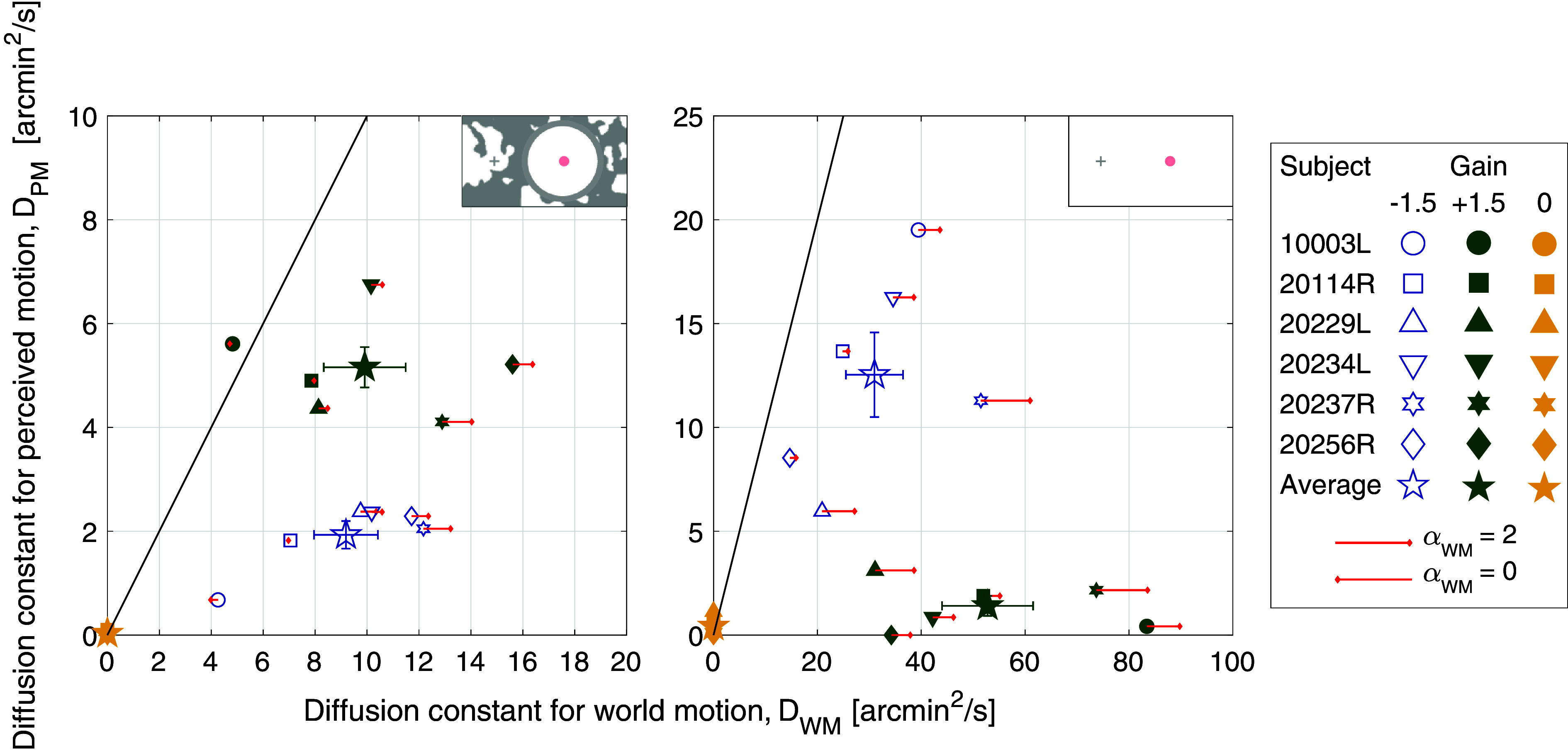
Detecting the motion of an object relative to a world-fixed frame of reference is an exquisite human capability [G. E. Legge, F. Campbell, Vis. Res. 21, 205-213 (1981)]. However, there is a special condition where humans are unable to accurately detect relative motion: Images moving in a direction consistent with retinal slip where the motion is unnaturally amplified can, under some conditions, appear stable [D. W. Arathorn, S. B. Stevenson, Q. Yang, P. Tiruveedhula, A. Roorda, J. Vis. 13, 22 (2013)]. We asked: Is world-fixed retinal image background content necessary for the visual system to compute the direction of eye motion, and consequently generate stable percepts of images moving with amplified slip? Or, are nonvisual cues sufficient? Subjects adjusted the parameters of a stimulus moving in a random trajectory to match the perceived motion of images moving contingent to the retina. Experiments were done with and without retinal image background content. The perceived motion of stimuli moving with amplified retinal slip was suppressed in the presence of a visible background; however, higher magnitudes of motion were perceived under conditions when there was none. Our results demonstrate that the presence of retinal image background content is essential for the visual system to compute its direction of motion. The visual content that might be thought to provide a strong frame of reference to detect amplified retinal slips, instead paradoxically drives the misperception of relative motion.

Keywords: adaptive optics; eye movements; motion perception.


Conflict of interest statement

Competing interests statement:P.T. and A.R. are co-inventors on US Patent #10130253, assigned to the University of California.

Figures

Fig. 1.
Fig. 1.
Display configuration. (A) The projector drew a fixation target and the surrounding 17° field displayed either white light or a binarized noise pattern. The AOSLO generated the stimuli positioned 2° temporal to the fovea (nasal field). These images were simultaneously projected onto the retina through a beamsplitter. (B) Rules for the three retina-contingent stimuli. Each panel shows an identical retinal trajectory indicated by the orange arrows. The purple arrows indicate the stimulus’ trajectory in the world. A Gain −1.5 stimulus moves with increased retinal slip, a Gain +1.5 stimulus moves in advance of retinal motion, and a Gain 0 stimulus is world-fixed. The dashed-brown arrows indicate the stimulus’ trajectory across the retina. A Gain −1.5 stimulus moves 2.5× more on the retina than a Gain 0 stimulus, and a Gain 0 stimulus moves 2× more on the retina than a Gain +1.5 stimulus. (C) Simulated trajectories showing uncorrelated, Brownian motion (Left) and positively correlated, persistent motion (Right). The white cross indicates the starting position for each trace. Both paths were generated from the same average step length, L = 0.6 arcminutes, and have similar speeds (S), yet have different diffusion constants (D) due to different degrees of persistence, indicated by the value α. Full descriptions of these parameters are in Materials and Methods. (D and E) Experiment sequence. Subjects fixated on the target and attended to the stimuli positioned 2° temporally. The retina-contingent stimulus moved contingent to each subject’s idiosyncratic fixational eye motion. The random walk stimulus moved independent of eye motion, with pregenerated random offsets. Under background-present conditions (D), the projector field displayed a binarized noise pattern which changed after each presentation and the fixation target remained on for the entire duration. Under background-absent conditions (E), a “Ganzfeld effect” was achieved by setting up a white paper with an aperture in front of the display permitting only the AOSLO and projector beams to enter the eye, shown in (A). LEDs were taped around the eye to illuminate the paper and the luminance was adjusted so that the subject saw only the stimuli in a white full-field surround. Owing to its proximity to the eye, the natural blur of the aperture rendered the transition between the display and luminance-matched paper invisible. The fixation target was timed to turn off when the stimuli were presented.
Fig. 2.
Fig. 2.
Average diffusion constants for perceived motion (DPM) versus diffusion constants for world motion (DWM) from six subjects. Experiments were tested under background-present and background-absent conditions, indicated by labels on the Top Right corner of each graph. The small symbols represent each subject’s average perceptual match for Gain −1.5 stimuli (blue open symbols), Gain +1.5 stimuli (green filled symbols), and Gain 0 stimuli (yellow filled symbols). The large stars are the group averages with SE of the mean bars. The red arrows show the extent to which the eye motion, and consequent retina-contingent stimulus’ world motion (αWM), deviated from Brownian. Arrows pointing Right indicate persistence (αWM > 1), arrows pointing Left indicate antipersistence (αWM < 1), and no arrow means that the motion was Brownian (αWM = 1 +/− 0.02). Longer arrows correspond to higher deviations from Brownian motion. The arrow length in the legend indicates pure persistence (αWM = 2, straight line trajectory at constant velocity) if pointing Right or pure antipersistence (αWM = 0, oscillatory motion) if pointing Left.
Fig. 3.
Fig. 3.
Computed ratios from six subjects tested under background-present (y-axis) and background-absent (x-axis) conditions. The symbols represent each subject’s [average diffusion constant for perceived motion]/[average diffusion constant for world motion] for two Gain conditions: Gain −1.5 stimuli (blue open symbols) and Gain +1.5 stimuli (green filled symbols). The red arrows show the extent to which the eye motion, and consequent retina-contingent stimulus’ world motion (αWM), deviated from Brownian. Arrows pointing up and Right indicate persistence (αWM > 1) under background-present and background-absent conditions, respectively. Arrows pointing down and Left indicate antipersistence (αWM < 1) under background-present and background-absent conditions, respectively. No arrow means that the motion was Brownian (αWM = 1 +/− 0.02). Longer arrows correspond to higher deviations from Brownian motion. The arrow length in the legend indicates pure persistence (αWM = 2, straight line trajectory at constant velocity) if pointing Up/Right or pure antipersistence (αWM = 0, oscillatory motion) if pointing Down/Left.
Fig. 4.
Fig. 4.
(AC) Violin plots (9) showing the distribution of the (A) diffusion constant for eye motion, DEM, (B) α for eye motion, αEM, and (C) speed for eye motion, SEM, for three Gains (−1.5, +1.5, and 0) under two conditions (background-present and background-absent). In the center of the violins, the white circles and dark bars represent the median and interquartile range, respectively; these are surrounded by the density trace (10). The black points are the (A) average DEM, (B) average αEM, and (C) average SEM for each subject across their six respective trials for each Gain. The asterisks indicate statistical significance (P < 0.05 and P < 0.01, respectively) from a post hoc Tukey–Kramer test following a two-factor repeated-measures ANOVA.
Fig. 5.
Fig. 5.
Example gaze traces for subject 10003L during Gain −1.5 retina-contingent presentations under (A) background-present (1,500-ms duration and αEM = 0.90) and (B) background-absent (750-ms duration and αEM = 1.29) conditions. The white cross indicates the starting position for each trace. The gaze directions are labeled: Left (L), Right (R), Up (U), and Down (D).
Fig. 6.
Fig. 6.
Computed ratios from six subjects plotted as a function of each subject’s α for world motion, αWM. The symbols represent each subject’s [average diffusion constant for perceived motion]/[average diffusion constant for world motion]. Data from two experiments are shown: Gain −1.5 stimuli tested under background-absent conditions (blue open symbols) and Gain +1.5 stimuli tested under background-present conditions (green filled symbols). The black curve predicts the DPM/DWM versus αWM, considering that the subject matched the mean square displacements of the random walk stimulus with the retina-contingent stimulus over a time interval of 2 frames. Because the random walk stimulus’ motion is Brownian (αPM = 1) and the retina-contingent stimulus’ motion ranges from antipersistent (0.9 <= αWM < 1), to Brownian (αWM = 1), to persistent (1 < αWM <= 1.8), the curve shows an exponential decay, which largely matches the data.

Update of

References

    1. Legge G. E., Campbell F., Displacement detection in human vision. Vis. Res. 21, 205–213 (1981). - PubMed
    1. Wang Y., et al. , Human foveal cone photoreceptor topography and its dependence on eye length. eLife 8, e47148 (2019). - PMC - PubMed
    1. Westheimer G., Visual acuity and hyperacuity. Invest. Ophthalmol. Vis. Sci. 14, 570–572 (1975). - PubMed
    1. McKee S. P., Welch L., Taylor D. G., Bowne S. F., Finding the common bond: Stereoacuity and the other hyperacuities. Vis. Res. 30, 879–891 (1990). - PubMed
    1. Riggs L. A., Ratliff F., Cornsweet J. C., Cornsweet T. N., The disappearance of steadily fixated visual test objects. J. Opt. Soc. Am. 43, 495–501 (1953). - PubMed

LinkOut - more resources