The diagnostic performance of cochlear endolymphatic hydrops and perilymphatic enhancement in stratifying Ménière's disease probabilities: A meta-analysis of semi-quantitative MRI-based grading systems
- PMID: 39570877
- PMCID: PMC11581247
- DOI: 10.1371/journal.pone.0310045
The diagnostic performance of cochlear endolymphatic hydrops and perilymphatic enhancement in stratifying Ménière's disease probabilities: A meta-analysis of semi-quantitative MRI-based grading systems
Abstract
Background: The diagnosis of Meniere's Disease (MD) presents significant challenges due to its complex symptomatology and the absence of definitive biomarkers. Advancements in MRI technology have spotlighted endolymphatic hydrops (EH) as a key pathological marker, necessitating a reevaluation of its diagnostic utility amidst the need for standardized and validated MRI-based grading scales.
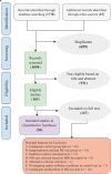
Methods: Our meta-analysis scrutinized the diagnostic efficacy of semi-quantitative MRI-based cochlear endolymphatic hydrops (EH) and perilymphatic enhancement (PLE) grading systems in delineating clinically relevant discriminations: "Spotting" the shift from normal or asymptomatic ears to possible/probable MD (pMD), "Confirming" the progression to definite MD (dMD), and "Establishing" the presence of dMD. A thorough literature search up to October 2023 resulted in 35 pertinent studies, forming the basis of our analysis through a bivariate mixed-effects regression model.
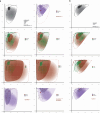
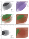
Results: Using criteria from the American Academy of Otolaryngology-Head and Neck Surgery (AAO-HNS) and Barany Society, across varying thresholds and disease probabilities; the Establishment model at an EH grade 1 threshold revealed a sensitivity of 85.4% and a specificity of 82.7%. Adjusting the threshold to EH grade 2 results in a sensitivity increase to 92.1% (CI: 85.9-95.7) and a specificity decrease to 70.6% (CI: 64.5-76.1), with a DOR of 28.056 (CI: 14.917-52.770). The Confirmation model yields a DOR of 5.216, indicating a lower diagnostic accuracy. The Spotting model demonstrates a sensitivity of 48.3% (CI: 34.8-62.1) and a specificity of 88.0% (CI: 77.8-93.9), with a DOR of 6.882. The normal ears subgroup demonstrated a notably high specificity of 89.7%, while employing Nakashima's criteria resulted in a reduced sensitivity of 74.9%, significantly diverging from other systems (p-value < 0.001). The PLE grading system showcased exceptional sensitivity of 98.4% (CI: 93.7-99.6, p-value < 0.001).
Conclusion: Our meta-analysis supports a tailored diagnostic approach for MD, emphasizing the need for effective grading systems at each stage. For "Spotting," the model shows high specificity but requires improved sensitivity, suggesting additional criteria are needed. The "Confirming" stage highlights the need for refined, sensitive grading systems due to lower diagnostic accuracy. In the "Establishing" stage, an EH grade 1 threshold is effective, but grade 2 enhances sensitivity while reducing specificity, indicating a need for balance. The PLE grading system excels in sensitivity, making it highly reliable. High specificity in the normal ears subgroup confirms accurate non-pathological distinction, though Nakashima's criteria show reduced sensitivity, underscoring variability in grading systems. These findings advocate for a standardized, unified grading system balancing sensitivity and specificity across all MD stages to optimize diagnostics and clinical outcomes.
Copyright: © 2024 Azarpey et al. This is an open access article distributed under the terms of the Creative Commons Attribution License, which permits unrestricted use, distribution, and reproduction in any medium, provided the original author and source are credited.
Conflict of interest statement
The authors have declared that no competing interests exist.
Figures


Similar articles
-
Delayed post gadolinium MRI descriptors for Meniere's disease: a systematic review and meta-analysis.Eur Radiol. 2023 Oct;33(10):7113-7135. doi: 10.1007/s00330-023-09651-8. Epub 2023 May 12. Eur Radiol. 2023. PMID: 37171493 Free PMC article.
-
The value of four stage vestibular hydrops grading and asymmetric perilymphatic enhancement in the diagnosis of Menière's disease on MRI.Neuroradiology. 2019 Apr;61(4):421-429. doi: 10.1007/s00234-019-02155-7. Epub 2019 Feb 5. Neuroradiology. 2019. PMID: 30719545 Free PMC article.
-
Correlation between magnetic resonance imaging classification of endolymphatic hydrops and clinical manifestations and audiovestibular test results in patients with definite Ménière's disease.Auris Nasus Larynx. 2022 Feb;49(1):34-45. doi: 10.1016/j.anl.2021.03.027. Epub 2021 Apr 15. Auris Nasus Larynx. 2022. PMID: 33865653
-
Correlation of endolymphatic hydrops and perilymphatic enhancement with the clinical features of Ménière's disease.Eur Radiol. 2024 Sep;34(9):6036-6046. doi: 10.1007/s00330-024-10620-y. Epub 2024 Feb 3. Eur Radiol. 2024. PMID: 38308680
-
Systematic review of magnetic resonance imaging for diagnosis of Meniere disease.J Vestib Res. 2019;29(2-3):121-129. doi: 10.3233/VES-180646. J Vestib Res. 2019. PMID: 31356219
References
-
- van Steekelenburg JM, van Weijnen A, de Pont LMH, Vijlbrief OD, Bommeljé CC, Koopman JP, et al.. Value of Endolymphatic Hydrops and Perilymph Signal Intensity in Suspected Ménière Disease. AJNR Am J Neuroradiol. 2020;41(3):529–34. Epub 2020/02/08. doi: 10.3174/ajnr.A6410 ; PubMed Central PMCID: PMC7077918. - DOI - PMC - PubMed
-
- Homann G, Vieth V, Weiss D, Nikolaou K, Heindel W, Notohamiprodjo M, et al.. Semi-quantitative vs. volumetric determination of endolymphatic space in Menière’s disease using endolymphatic hydrops 3T-HR-MRI after intravenous gadolinium injection. PLoS One. 2015;10(3):e0120357. Epub 2015/03/15. doi: 10.1371/journal.pone.0120357 ; PubMed Central PMCID: PMC4358992. - DOI - PMC - PubMed
-
- Pyykkö I, Nakashima T, Yoshida T, Zou J, Naganawa S. Meniere’s disease: a reappraisal supported by a variable latency of symptoms and the MRI visualisation of endolymphatic hydrops. BMJ Open. 2013;3(2). Epub 2013/02/19. doi: 10.1136/bmjopen-2012-001555 ; PubMed Central PMCID: PMC3586172. - DOI - PMC - PubMed
Publication types
MeSH terms
LinkOut - more resources
Full Text Sources
Medical

