. 2024 Nov 21;20(11):e1012741.
doi: 10.1371/journal.ppat.1012741. eCollection 2024 Nov.

SARS-CoV-2 evolution balances conflicting roles of N protein phosphorylation

Affiliations

SARS-CoV-2 evolution balances conflicting roles of N protein phosphorylation

Abdullah M Syed et al. PLoS Pathog. .

Abstract

All lineages of SARS-CoV-2, the coronavirus responsible for the COVID-19 pandemic, contain mutations between amino acids 199 and 205 in the nucleocapsid (N) protein that are associated with increased infectivity. The effects of these mutations have been difficult to determine because N protein contributes to both viral replication and viral particle assembly during infection. Here, we used single-cycle infection and virus-like particle assays to show that N protein phosphorylation has opposing effects on viral assembly and genome replication. Ancestral SARS-CoV-2 N protein is densely phosphorylated, leading to higher levels of genome replication but 10-fold lower particle assembly compared to evolved variants with low N protein phosphorylation, such as Delta (N:R203M), Iota (N:S202R), and B.1.2 (N:P199L). A new open reading frame encoding a truncated N protein called N*, which occurs in the B.1.1 lineage and subsequent lineages of the Alpha, Gamma, and Omicron variants, supports high levels of both assembly and replication. Our findings help explain the enhanced fitness of viral variants of concern and a potential avenue for continued viral selection.


Conflict of interest statement

AMS and JAD are co-inventors on a patent application filed by Gladstone Institutes and University of California on the generation of SARS-CoV-2 VLPs. TYT and MO are inventors on a patent application filed by the Gladstone Institutes that covers the use of pGLUE to generate SARS-CoV-2 infectious clones and replicons. The NJK Laboratory has received research support from Vir Biotechnology, F. Hoffmann-La Roche, and Rezo Therapeutics. NJK has a financially compensated consulting agreement with Maze Therapeutics. NJK is the President and is on the Board of Directors of Rezo Therapeutics, and he is a shareholder in Tenaya Therapeutics, Maze Therapeutics, Rezo Therapeutics, GEn1E Lifesciences, and Interline Therapeutics. All other authors declare no competing interests.

Figures
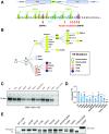
Fig 1
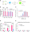
Fig 1. SARS-CoV-2 variants have reduced N protein phosphorylation.
A) Map of SARS-CoV-2 N protein showing phosphorylation sites within the serine/arginine rich motif (SR). Proposed phosphorylation sites and kinases indicated with arrows. Location of common mutations found in circulating SARS-CoV-2 lineages indicated with red arrows (P199L, S202R, R203M, R203K, G204R, T205I). B) Schematic illustration of the phylogeny of circulating variants with predominant SR mutations indicated by color. C) Autoradiography (32P) and quantification (D) of purified N variants after in vitro phosphorylation with SRPK, GSK3 and CK1 using 32P-γ-ATP (see S1A Fig). The blot is representative of 3 independent experiments and the quantification represents 3 independent experiments shown as mean +/- SD normalized to WT. *, p<0.05; **, p<0.01; ***, p<0.001; ****, p<0.0001 by Student’s T-test compared to the WT control. E) SDS-PAGE gel of immunoprecipitated strep-tagged N-protein variants isolated from transfected 293T cells. Gel visualized by Krypton staining (see S1D Fig).
Fig 2
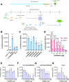
Fig 2. N protein phosphorylation inhibits virus-like particle assembly.
A) Process flow for generating and testing SARS-CoV-2 virus-like particles packaging luciferase (VLPs). B) Normalized luminescence signal induced by VLPs generated using six variant N proteins with and without treatment with SB216763, a GSK3 inhibitor, at 10 μM. P199L, S202R and R203M have previously been shown to enhance VLP assembly [1] while G204R and M234I have similar packaging efficiency as the ancestral protein. The data are presented as mean +/- SD of three independent experiments. P values are indicated for piecewise comparisons using Student’s T-test. C) VLP-induced signal generated using SARS-CoV-2 or SARS-CoV-1 N protein incubated with varying concentrations of CHIR 98014, another GSK3 inhibitor. The data are presented as mean +/- SD of four independent experiments. P values are indicated for piecewise comparisons using Student’s T-test. D and E) Normalized luminescence signal from VLPs generated using N variants with alanine or glutamic acid substitutions at (D) critical GSK3 phosphorylation sites (198, 202, 206) or (E) neighboring sites (197, 201, 205). The data are presented as mean +/- SD of four independent experiments. P values are indicated for piecewise comparisons using Student’s T-test.
Fig 3
Fig 3. N protein phosphorylation enhances single cycle infection.
A) Design of SARS-CoV-2 N-deleted replicon and process flow for single cycle infection. Clip art was created with BioRender.com with permission. B) and C) Luminescence from 293T-ACE2/TMPRSS2 cells transfected with packaging defective N mutants and infected with N-deleted single cycle infectious particles (ΔN-SCIPs). Pax indicates treatment with 3 μM nirmatrelvir. D) Luminescence from cells transfected with packaging defective ancestral N protein or a control plasmid and treated with varying concentrations of CHIR98014, a GSK3 inhibitor. E-H) Luminescence from cells infected with nanoluciferase expressing ΔS replicon encoding ancestral N sequence and treated with varying concentrations of CHIR98014. All conditions with CHIR98014 or nirmatrelvir treatment were pre-treated 24 hours prior to infection, during infection and post-infection at the same concentration. Vero-ACE2/TMPRSS2 cells were additionally treated with 2μM CP-100356 (Millipore Sigma) to inhibit the P-glycoprotein efflux pump overexpressed in this cell line. For B-H, the data are presented as mean +/- SD of four (B-G) or three (H) independent experiments. *, p<0.05; **, p<0.01; ***, p<0.001; ****, p<0.0001 by Student’s T-test.
Fig 4
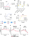
Fig 4. N protein phosphorylation inhibits viral fitness.
A) Luminescence from cells infected with WA1 and Delta infectious clones with NanoLuc luciferase inserted in place of Orf7a and Orf7b. Cells were treated with varying concentrations of CHIR98014 as indicated. WA1 contains ancestral N protein while Delta contains the R203M substitution which reduces N protein phosphorylation. The data are presented as mean +/- SD of three independent experiments. *, p<0.05; **, p<0.01; ***, p<0.001 for piecewise comparisons conducted by Student’s T-test. B) Plaque forming units of infectious clones encoding ancestral (WT), S202R or R203M mutations within the N protein, 72 hours post infection in A549-ACE2, Vero TMPRSS2, Calu-3 and Huh7.5 ACE2/TMPRSS2 cells. The data are presented as mean +/- SD of three (A549-ACE2, Vero TMPRSS2, and Calu-3) or two (Huh7.5 ACE2/TMPRSS2) independent experiments conducted in triplicate. P values from Student’s T-test are indicated in the panel. C) Schematic of viral competition assay. Clip art was created with BioRender.com with permission. D) Quantification of a pilot competition experiment between R203M mutant virus on WA1 background using Sanger sequencing after co-infection with 1:1, 10:1, or 100:1 ratios of WA1:R203M on A549-ACE2 cells. The data is presented as % of mutant based on Sanger sequencing from a single biological experiment. E-G) Quantification of competition between mutant viruses (R203M, S188A/S206A and M234I) on WA1 background relative to the WA1 quantified through qPCR. Gray lines indicate results of competition in untreated cells while red lines indicate treatment with CHIR98014 at 2.5 μM. The data are presented as mean +/- SD of three independent experiments.
Fig 5
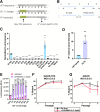
Fig 5. N* improves viral fitness independent of phosphorylation.
A) Annotation of the 28881–28893: ggg>aac mutation showing the generation of the “ACGAAC” TRS sequence. B) Schematic of truncated N proteins produced to test effects on assembly. C) Luminescence from VLP treated 293T-ACE2/TMPRSS2 cells. VLPs were generated with indicated truncated N proteins. Only N* (N:210–419) is competent for delivering RNA. The data are presented as mean +/- SD of one independent experiment conducted in six replicates. ***, p<0.001; ****, p<0.0001 by Student’s T-test compared to wildtype N. D) Quantification of Western blot (streptactin-HRP) from producer cell lysate and purified VLPs showing relative N* abundance compared to full length N protein. Both proteins contain a C-terminal 2xStrep-Tag. The data are presented as mean +/- SD of one independent experiment conducted in triplicate and the blot is shown in S4 Fig. **, p<0.01 by Student’s T-test. E) Luminescence from VLP treated cells with VLPs generated with combinations of N* and full length N protein and with or without treatment with CHIR98014. The data are presented as mean +/- SD of one independent experiment conducted in six replicates. P values are indicated for piecewise comparisons conducted with Student’s T-test. F, G) Viral competition assays of G204R-TRS virus (expresses N*) and G204R virus (no N*) with WA1 infectious clone with or without CHIR98014 treatment as measured by qPCR. The data are presented as mean +/- SD of three independent experiments.

References

    1. Syed AM, Taha TY, Tabata T, Chen IP, Ciling A, Khalid MM, et al.. Rapid assessment of SARS-CoV-2-evolved variants using virus-like particles. Science. 2021;374: 1626–1632. doi: 10.1126/science.abl6184 - DOI - PMC - PubMed
    1. Wu H, Xing N, Meng K, Fu B, Xue W, Dong P, et al.. Nucleocapsid mutations R203K/G204R increase the infectivity, fitness, and virulence of SARS-CoV-2. Cell Host Microbe. 2021;29: 1788–1801.e6. doi: 10.1016/j.chom.2021.11.005 - DOI - PMC - PubMed
    1. Johnson BA, Zhou Y, Lokugamage KG, Vu MN, Bopp N, Crocquet-Valdes PA, et al.. Nucleocapsid mutations in SARS-CoV-2 augment replication and pathogenesis. PLoS Pathog. 2022;18: e1010627. doi: 10.1371/journal.ppat.1010627 - DOI - PMC - PubMed
    1. Yaron TM, Heaton BE, Levy TM, Johnson JL, Jordan TX, Cohen BM, et al.. Host protein kinases required for SARS-CoV-2 nucleocapsid phosphorylation and viral replication. Sci Signal. 2022;15: eabm0808. doi: 10.1126/scisignal.abm0808 - DOI - PMC - PubMed
    1. Liu X, Verma A, Garcia G Jr, Ramage H, Lucas A, Myers RL, et al.. Targeting the coronavirus nucleocapsid protein through GSK-3 inhibition. Proc Natl Acad Sci U S A. 2021;118. doi: 10.1073/pnas.2113401118 - DOI - PMC - PubMed

Substances

Supplementary concepts