Review
. 2025 Jan 7;39(1-2):109-131.
doi: 10.1101/gad.352084.124.

mTORC1, the maestro of cell metabolism and growth

Affiliations
Review

mTORC1, the maestro of cell metabolism and growth

Long He et al. Genes Dev. .

Abstract

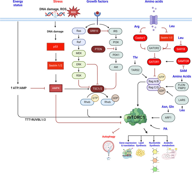
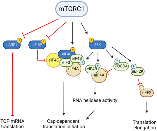
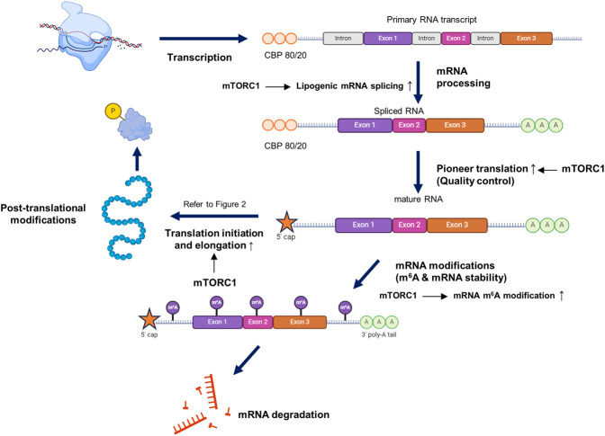
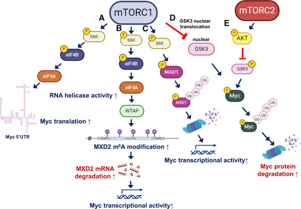
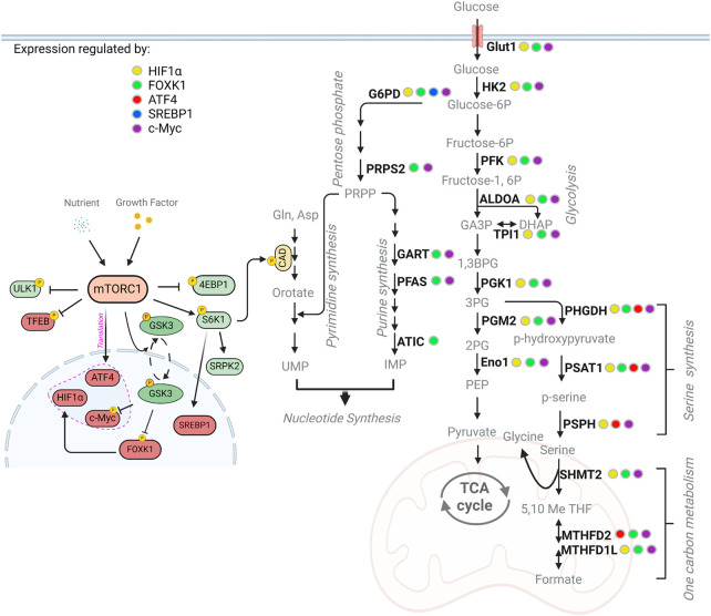
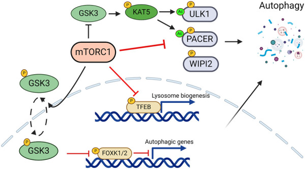
The mechanistic target of rapamycin (mTOR) pathway senses and integrates various environmental and intracellular cues to regulate cell growth and proliferation. As a key conductor of the balance between anabolic and catabolic processes, mTOR complex 1 (mTORC1) orchestrates the symphonic regulation of glycolysis, nucleic acid and lipid metabolism, protein translation and degradation, and gene expression. Dysregulation of the mTOR pathway is linked to numerous human diseases, including cancer, neurodegenerative disorders, obesity, diabetes, and aging. This review provides an in-depth understanding of how nutrients and growth signals are coordinated to influence mTOR signaling and the extensive metabolic rewiring under its command. Additionally, we discuss the use of mTORC1 inhibitors in various aging-associated metabolic diseases and the current and future potential for targeting mTOR in clinical settings. By deciphering the complex landscape of mTORC1 signaling, this review aims to inform novel therapeutic strategies and provide a road map for future research endeavors in this dynamic and rapidly evolving field.

Keywords: cancer; cellular signaling; mT; mTOR complex 1; mTORC1.


Figures

Figure 1.
Figure 1.
Major upstream regulators of mTORC1. Growth factors, amino acids, energy levels, phosphatidic acid (PA), and cellular stresses, including DNA damage and reactive oxygen species (ROS), modulate mTORC1 activity, which in turn regulates numerous biological processes, including gene expression, nucleotide synthesis, protein translation, lipid synthesis, and autophagy. Negative regulators of mTORC1 are highlighted in red. (Figure created with BioRender.com.)
Figure 2.
Figure 2.
Translation regulation by mTORC1 signaling. mTORC1 promotes translation of terminal oligopyrimidine (TOP) mRNAs via LARP1 phosphorylation. mTORC1 also enhances cap-dependent translation initiation through phosphorylation of 4E-BP and components of the translation initiation complex. S6K stimulates translation initiation by phosphorylating eIF4B, which promotes its recruitment into the translation preinitiation complex while also phosphorylating PDCD4 and inducing its ubiquitin-mediated turnover, thereby boosting eIF4A RNA helicase activity. S6K also promotes translation elongation through phosphorylation of eEF2K. (Figure created with BioRender.com.)
Figure 3.
Figure 3.
mTORC1–S6K and the central dogma. mTORC1–S6K modulates multiple stages of gene expression, including mRNA transcription, processing (such as mRNA splicing), the pioneer round of translation for quality control, and post-transcriptional (m6A) and post-translational modifications. For instance, mRNA splicing of lipogenic mRNA, the pioneer round of translation, and m6A modification are promoted by mTORC1 activation. This regulation ensures precise control of gene and protein expression. (Figure created with BioRender.com.)
Figure 4.
Figure 4.
The complex regulation of cMyc downstream from mTORC1. (A) Upregulation of cMyc translation via increased eIF4A RNA helicase activity through the eIF4B–S6K pathway to resolve highly structured 5′ UTRs of Myc mRNA. (B) Enhanced cMyc activity through destabilization of MXD2 mRNA by mTORC1-mediated m6A modification. (C) Increased cMyc activity through the degradation of MAD1, the negative regulator of Myc, via mTORC1–S6K-mediated phosphorylation. (D) Induction of cMyc stability through the suppression of GSK3 nuclear translocation by mTORC1. (E) Growth factor or nutrient activation of AKT suppresses GSK3-mediated cMyc degradation through GSK3 phosphorylation. (Figure created with BioRender.com.)
Figure 5.
Figure 5.
The mTORC1 signaling pathway extensively controls cellular metabolism. This schematic illustrates the metabolic genes induced by mTORC1 within their respective pathways, based on our current understanding of mTORC1's regulation of glycolysis, the pentose phosphate pathway (PPP), serine/one carbon metabolism, and nucleotide metabolism. mTORC1 orchestrates this metabolic rewiring by regulating gene expression through multiple transcription factors, including Hif1a (yellow), FOXK (green), ATF4 (red), and SREBP (blue). (Figure created with BioRender.com.)
Figure 6.
Figure 6.
mTORC1 regulation of autophagy. mTORC1 regulates autophagy through both transcriptional and post-translational mechanisms. (Figure created with BioRender.com.)

Similar articles

Cited by

References

    1. Adams CM. 2007. Role of the transcription factor ATF4 in the anabolic actions of insulin and the anti-anabolic actions of glucocorticoids. J Biol Chem 282: 16744–16753. 10.1074/jbc.M610510200 - DOI - PubMed
    1. Alesi N, Akl EW, Khabibullin D, Liu HJ, Nidhiry AS, Garner ER, Filippakis H, Lam HC, Shi W, Viswanathan SR, et al. 2021. TSC2 regulates lysosome biogenesis via a non-canonical RAGC and TFEB-dependent mechanism. Nat Commun 12: 4245. 10.1038/s41467-021-24499-6 - DOI - PMC - PubMed
    1. Alessi DR, Kozlowski MT, Weng QP, Morrice N, Avruch J. 1998. 3-Phosphoinositide-dependent protein kinase 1 (PDK1) phosphorylates and activates the p70 S6 kinase in vivo and in vitro. Curr Biol 8: 69–81. 10.1016/S0960-9822(98)70037-5 - DOI - PubMed
    1. Angarola B, Ferguson SM. 2019. Weak membrane interactions allow Rheb to activate mTORC1 signaling without major lysosome enrichment. Mol Biol Cell 30: 2750–2760. 10.1091/mbc.E19-03-0146 - DOI - PMC - PubMed
    1. Aranovich A, Hua R, Rutenberg AD, Kim PK. 2014. PEX16 contributes to peroxisome maintenance by constantly trafficking PEX3 via the ER. J Cell Sci 127: 3675–3686. 10.1242/jcs.146282 - DOI - PMC - PubMed

Substances

LinkOut - more resources