. 2024 Nov 21;15(1):10100.
doi: 10.1038/s41467-024-54438-0.

Multimodal single cell-resolved spatial proteomics reveal pancreatic tumor heterogeneity

Affiliations

Multimodal single cell-resolved spatial proteomics reveal pancreatic tumor heterogeneity

Yanfen Xu et al. Nat Commun. .

Abstract

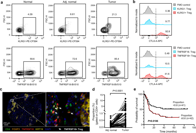
Despite the advances in antibody-guided cell typing and mass spectrometry-based proteomics, their integration is hindered by challenges for processing rare cells in the heterogeneous tissue context. Here, we introduce Spatial and Cell-type Proteomics (SCPro), which combines multiplexed imaging and flow cytometry with ion exchange-based protein aggregation capture technology to characterize spatial proteome heterogeneity with single-cell resolution. The SCPro is employed to explore the pancreatic tumor microenvironment and reveals the spatial alternations of over 5000 proteins by automatically dissecting up to 100 single cells guided by multi-color imaging of centimeter-scale formalin-fixed, paraffin-embedded tissue slide. To enhance cell-type resolution, we characterize the proteome of 14 different cell types by sorting up to 1000 cells from the same tumor, which allows us to deconvolute the spatial distribution of immune cell subtypes and leads to the discovery of subtypes of regulatory T cells. Together, the SCPro provides a multimodal spatial proteomics approach for profiling tissue proteome heterogeneity.


Conflict of interest statement

Competing interests: R.T. is the founder of BayOmics, Inc. The other authors declare no competing interests.

Figures

Fig. 1
Fig. 1. Concept and workflow of the SCPro platform.
The SCPro platform integrates multiple modules. a Antibody-guided cell typing based on high-quality multiplexed imaging and automated LMD with single-cell resolution. b Flow cytometry-based cell typing. c Ultra-high-sensitivity proteomics platform combining with ion exchange-based protein aggregation capture sample preparation, low-flow chromatography, and high-sensitivity mass spectrometry data acquisition. d Decoding the pancreatic tumor microenvironment through spatial deconvolution. CAF cancer-associated fibroblast, iCAF inflammatory CAF, myCAF myofibroblastic CAF, apCAF antigen-presenting CAF, Treg regulatory T cell, FFPE formalin-fixed paraffin-embedded, mIHC multiplexed immunohistochemical, LMD laser microdissection, TME tumor microenvironment.
Fig. 2
Fig. 2. Development of iPAC technology for processing rare stained tissue cells.
a Workflow and principle of iPAC technology. b Identified protein groups and unique peptides from processing nanogram HEK 293T cell lysate or direct injection of an equal number of pre-digested HEK 293T peptides, showed in red and blue lines, respectively. The proportion shown on the graph indicates the protein recovery rate for each group. c Identified protein groups and unique peptides from 10 to 1000 flow cytometry-sorted HEK 293T cells. d Identified protein groups and unique peptides from 20, 50, and 100 μm-side length squares of 12 μm-thick H&E-stained mouse brain slice. The images shown are representative of 3 independent experiments. e Identified protein groups and unique peptides of 200 μm-side length squares from a 12 μm-thick H&E-stained mouse brain without (W/o) or with (W/) extended wash. The insert violin diagram shows the Coefficient of Variation (CV) distributions (n = 3 biological replicates). The insert boxplots display the median (white dot), the 25th and 75th percentiles (black box), and the minimum and maximum (whiskers). f Upper panel, Contamination Ratios (CR) of H&E-stained mouse brain samples W/o or W/ extended wash along the LC gradient. The pre-digested HeLa was served as a control. The transparent shades beyond each dot-line indicate the half standard deviation of CR within each group. The bottom panel identified peptide-spectrum matches (PSMs) along with retention time (RT). The total ion chromatogram (TIC) intensity of precursors extracted for generating heatmaps is displayed. g Representing heatmap of precursors in the trapped ion mobility (IM)-m/z space at specific RT from one of the three replicates in each group. h Workflow showing the acinar, tumor, and lymph regions (n = 4 biological replicates from one KPf/fC mouse). The images shown are all of 4 independent experiments. Scale bar, 2 mm and 500 μm for the left and right images, respectively. i Identified protein groups and peptides from different regions. j Principal Component Analysis (PCA) analysis based on quantified protein groups before data filtering. DDM N-dodecyl-β-D-maltoside, SAX strong anion exchange, ACN acetonitrile, ZDV zero-dead-volume. Source data are provided as a Source Data file.
Fig. 3
Fig. 3. mIHC profiling and LMD capture of single cells in the pancreatic TME.
a Spatial proteomics workflow of the SCPro. b Multiplexed immunohistochemical whole-slide image of a 4-μm-thick KPf/fC mouse tissue section. The color dot showing the representative LMD cutting position of distinct cell types (n = 3 biological replicates). Scale bar, 1 mm. c Representative tissue cytometry charts of the PDAC-1 region. d Workflow of the ROI selection, cell typing and cell mask generation, and image alignment based on the real-time image of LMD. Scale bar, 300 μm, 20 μm, 300 μm, 100 μm, and 20 μm for the images from left to right, respectively. e Automated LMD with single-cell resolution. The images shown are representative of 3 independent experiments. Scale bar, 10 μm. EpCAM (epithelial cells); CD45 (immune cells); αSMA (fibroblasts); DAPI (nuclei). PanIN pancreatic intraepithelial neoplasm, PDAC pancreatic ductal adenocarcinoma, ROI region of interest.
Fig. 4
Fig. 4. Uncovering the spatial proteomic heterogeneity of PDAC TME.
a Quantified protein groups of each cell type. A total area of 120,000 μm3 cell contours (~60 cells) for the CAF and IT regions, and 200,000 μm3 cell contours (~100 cells) for the Acinar, PanIN, PDAC, and LN regions were dissected from one KPf/fC mouse tissue section with 4-μm-thick (n = 3 biological replicates from one KPf/fC mouse). Boxplots display the median (horizontal line), the 25th and 75th percentiles (colored box), and the minimum and maximum (whiskers). b PCA analysis based on quantified protein groups before data filtering (n = 3 biological replicates from one KPf/fC mouse). Replicates of each subtype were wrapped in a colored circle. c Unsupervised hierarchical clustering of significantly expressed proteins in acinar, PanIN, and PDAC regions (P-value < 0.05, fold change >2). P-value was calculated using one-way ANOVA. Known cell-type-specific markers were labeled on the right. Protein expression levels were Z-scored. d Dot plot showing the Gene Ontology Biology Process (GOBP) in terms of significantly differentially expressed proteins. Significance was calculated by one-tailed Fisher’s Exact Test (P-value < 0.05). e Prognostic markers for pancreatic cancer identified in the dataset and classified by their functions (P-value < 0.05, fold change >1.2). P-value was calculated using one-way ANOVA. *, reported in other cancers as a prognostic marker; **, reported in pancreatic cancer as a prognostic marker. f PCA analysis based on quantified protein groups before data filtering. Replicates of each region were wrapped in a colored circle (n = 3 biological replicates from one KPf/fC mouse). g Unsupervised hierarchical clustering of significantly differential proteins for IT and LN regions (two-tailed Student’s t-test, P-value < 0.05, fold change >2). Known cell-type-specific markers were labeled on the right. Protein expression levels were Z-scored. h Dot plot showing the GOBP terms of significantly differentially expressed proteins. Significance was calculated by one-tailed Fisher’s Exact Test (P-value < 0.05). No adjustment was made for multiple comparisons in (c, d, e and h). Source data are provided as a Source Data file.
Fig. 5
Fig. 5. SCPro dissects the PDAC TME through spatial deconvolution.
a Cell number of each cell type. b Proteome coverage of each cell type, the identified and quantified protein groups are indicated in gray and dark, respectively (n = 4 and 5 biological replicates for apCAF and the other groups, respectively, from 5 KPf/fC mice). c PCA plot. Ellipses cover the four cell lineages indicated by colors. d Heatmap showing significantly differentially expressed proteins for 14 cell types with P-value < 0.05 and fold change >2. P-values were calculated using the moderated t-statistic (with a two-tailed test) from the LIMMA package, no adjustment was made for multiple comparisons. The well-known lineage markers were labeled on the right. Protein expression levels were Z-scored. e Dot plot showing the significantly enriched GOBP terms of cell-lineage-specific expressed proteins (n = 4 and 5 biological replicates for apCAF and the other groups, respectively, from 5 KPf/fC mice). Significance was calculated by one-tailed Fisher’s Exact Test (P-value < 0.05), and no adjustment was made for multiple comparisons. f Predicted proportion of the 14-flow cytometry-sorted immune cell subtypes in the IT and LN regions. g Boxplots showing the predicted proportion of CD11b+ myeloid cells (MYE, MAC, MO, and NUE) and CD3+ lymphoid cells (T4, Treg, and T8) in the IT and LN regions (n = 3 biological replicates from one KPf/fC mouse tissue section). Boxplots display the mean (horizontal line), and the 25th and 75th percentiles (bounds of box). The sum of the predicted proportions of the two cell types was set to 100% through normalization. h mIHC staining of CD11b+ myeloid cells and CD3+ lymphoid cells. The sum of the proportion of the two cell types was set to 100% through normalization. The images shown are representative of 2 independent experiments. Scale bar, 2 mm and 100 μm for the left and right images, respectively. Krt19 (epithelial cells); CD11b (myeloid cells); CD3 (T cells); DAPI (nuclei). PCC pancreatic cancer cell, MYE myeloid cell, NEU neutrophil, MO monocyte, MAC macrophage, DC dendritic cell. Source data are provided as a Source Data file.
Fig. 6
Fig. 6. Sub-cell types discovery by SCPro.
a Workflow for discovering functional sub-cell types. b Bar plot showing the number of PM proteins. The line chart showing their proportion in all identified protein groups for individual cell types. c Heatmap showing the significantly differentially expressed PM proteins (P-value < 0.05, fold change >2). P-values were calculated using the moderated t-statistic (with a two-tailed test) from the LIMMA package. The numbers on the left and right of brackets represent the number of significantly differentially expressed PM proteins and the total number of PM proteins for each cell type, respectively. Protein expression levels were Z-scored. d Line plot showing the scaled expression levels of significant proteins within four representative cell types (PCC, CAF, Treg, and DC), the top 2 of which are colored, respectively. e t-SNE plot showing the expression patterns of Tnfrsf18 and Klrg1 on CD25+ Treg cells. f Proteome comparison of Klrg1 Treg and Klrg1+ Treg. The significant proteins were shown in blue and red color, respectively (P-value < 0.05, fold change >2). P-values were calculated using the moderated t-statistic (with a two-tailed test) from the LIMMA package. g Dot plot showing the enriched GOBP terms of the Klrg1 Treg and Klrg1+ Treg (n = 3 biological replicates from 3 KPf/fC mice). Significance was calculated by one-tailed Fisher’s Exact Test (P-value < 0.05), and subsequently q-values were estimated to control the false discovery rate across multiple comparisons. h Bar plot showing the proportion of CTLA-4+ and CD69+ cells in Klrg1 and Klrg1+ Treg cells, respectively (n = 5 biological replicates from 5 KPf/fC mice). Significance was calculated by two-tailed Student’s t-test and data are presented as mean ± SD. i Predicted proportion of Klrg1+ Treg among the 8 Klrg1-associated cell types in the IT and LN regions, respectively (n = 3 biological replicates from one KPf/fC mouse). Boxplots display the mean (horizontal line), and the 25th and 75th percentiles (bounds of box). j Summary of discovering a Treg subtype and predicting its spatial location. Source data are provided as a Source Data file.
Fig. 7
Fig. 7. Validation of the Treg subtypes in human PDAC samples.
a Scatter plot showing the expression levels of KLRG1 and TNFRSF18 in the Treg cells of human PDAC samples from the same patient (n = 1). Normal, pancreatic tissues located distant from the tumor; Adj. normal, adjacent normal samples near the tumor; Tumor, tumor sample. b Histograms with corresponding percentages showing the expression levels of CTLA-4 in the FMO control, KLRG1 Treg, KLRG1+ Treg, TNFRSF18 Treg, and TNFRSF18+ Treg, respectively. c Representative images of TNFRSF18+ Treg (CD4+FOXP3+TNFRSF18+ cells) in the tumor sample of human PDAC tissue microarray (TMA) slice using mIHC. Images shown are representative of 83 pairs of adjacent normal and tumor samples on the TMA slide. Scale bar, 200 μm and 10 μm for the left and right images, respectively. d Proportion of TNFRSF18+ Treg among CD4+ T cells in the PDAC TMA slide using mIHC (n = 81 pairs of adjacent normal and tumor samples). Significance was calculated by a two-tailed Student’s t-test. e Survival analysis of TNFRSF18+ Treg in PDAC TMA cohort (n = 83 PDAC tumor samples). The median proportion of TNFRSF18+ Treg was used as the cutoff to define the low and high-expression groups. P-values were determined by log-rank test. For sample information, see Supplementary Data 5. Source data are provided as a Source Data file.

References

    1. Mund, A., Brunner, A. D. & Mann, M. Unbiased spatial proteomics with single-cell resolution in tissues. Mol. Cell82, 2335–2349 (2022). - PubMed
    1. Mao, Y., Wang, X., Huang, P. & Tian, R. Spatial proteomics for understanding the tissue microenvironment. Analyst146, 3777–3798 (2021). - PubMed
    1. Wagner, J. et al. A single-cell atlas of the tumor and immune ecosystem of human breast cancer. Cell177, 1330–1345.e1318 (2019). - PMC - PubMed
    1. Giesen, C. et al. Highly multiplexed imaging of tumor tissues with subcellular resolution by mass cytometry. Nat. Methods11, 417–422 (2014). - PubMed
    1. Schurch, C. M. et al. Coordinated cellular neighborhoods orchestrate antitumoral immunity at the colorectal cancer invasive front. Cell182, 1341–1359.e1319 (2020). - PMC - PubMed

Publication types

MeSH terms

Associated data

LinkOut - more resources