. 2024 Nov 21;15(1):10106.
doi: 10.1038/s41467-024-54480-y.

Functional annotation of the Hippo pathway somatic mutations in human cancers

Affiliations

Functional annotation of the Hippo pathway somatic mutations in human cancers

Han Han et al. Nat Commun. .

Abstract

The Hippo pathway is commonly altered in cancer initiation and progression; however, exactly how this pathway becomes dysregulated to promote human cancer development remains unclear. Here we analyze the Hippo somatic mutations in the human cancer genome and functionally annotate their roles in targeting the Hippo pathway. We identify a total of 85 loss-of-function (LOF) missense mutations for Hippo pathway genes and elucidate their underlying mechanisms. Interestingly, we reveal zinc-finger domain as an integral structure for MOB1 function, whose LOF mutations in head and neck cancer promote tumor growth. Moreover, the schwannoma/meningioma-derived NF2 LOF mutations not only inhibit its tumor suppressive function in the Hippo pathway, but also gain an oncogenic role for NF2 by activating the VANGL-JNK pathway. Collectively, our study not only offers a rich somatic mutation resource for investigating the Hippo pathway in human cancers, but also provides a molecular basis for Hippo-based cancer therapy.


Conflict of interest statement

Competing interests: K.-L.G. is a co-founder of and holds an equity interest in Vivace Therapeutics. The other authors declare no competing interests.

Figures

Fig. 1
Fig. 1. Analysis of Hippo signaling gene alterations in the human cancer genome.
a Summary of Hippo signaling gene alterations in TCGA. Information of the TCGA patient samples was obtained from cBioportal. Hippo signaling genes refer to the Hippo pathway core components, effectors YAP and TAZ, and transcription factors TEAD1-4. b Around 1500 cancer patient samples carry multiple altered Hippo signaling genes. ce Histological analysis of the cancer patient samples with the altered Hippo signaling genes. Tumor samples were analyzed for their neoplasm histologic grade (c), tumor stage (d), and neoplasm disease stage (e) using chi-squared test. f Summary of the alteration types for Hippo signaling genes. Hippo signaling genes were analyzed for their amplification, deletion, mutation, and multi-alterations (i.e., mutation occurs together with either amplification or deletion) in TCGA patient samples. g Overview of the Hippo signaling gene alterations across 32 cancer types in TCGA. Total patient sample number for each cancer type was indicated. h Illustration of the Hippo signaling gene somatic mutations in TCGA. The number of patient samples carrying the somatic mutations was indicated for each Hippo signaling gene. Source data are provided as a Source Data file.
Fig. 2
Fig. 2. Functional annotation of the Hippo pathway somatic mutations.
a Illustration of the workflow for characterizing Hippo pathway missense mutations. The TCGA-documented Hippo pathway missense mutants were generated and reconstituted into their corresponding KO cells. The total missense mutation number for each Hippo pathway gene was indicated. b, c Summary of the identified LOF (b) and neutral (c) missense mutations for each Hippo pathway gene. d, e Summary of the cancer patients carrying the LOF (d) and neutral (e) missense mutations for each Hippo pathway gene. f Summary of the cancer patients carrying the Hippo LOF missense mutations across different cancer types. For each cancer type, the numbers of patients carrying the LOF and total missense mutations of the Hippo pathway genes were indicated. The Hippo LOF missense mutation rate for each cancer type was shown as a heatmap. The information of NF2 missense mutations in schwannoma and meningioma was obtained from COSMIC. g Summary of the LOF missense mutations identified for each Hippo pathway gene. The cancer type and mutation number were indicated. Source data are provided as a Source Data file.
Fig. 3
Fig. 3. Characterization of the LATS1/2 LOF missense mutations.
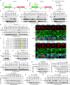
a, b Illustration of the protein domains and the identified LOF missense mutations for LATS1/2. c Illustration of the in vitro kinase assay used for validating the LATS1/2 LOF missense mutations. The purified LATS1/2 LOF missense mutants were subjected to in vitro kinase assay using bacterially purified GST-YAP as substrate. d, e The LATS1 LOF missense mutations inhibit LATS1 activity. The SFB-tagged LATS1 MOB1-binding domain (MBD)-associated LOF mutants (d) and its kinase domain-associated LOF mutants (e) were expressed in HEK293T cells, purified using S protein beads, washed thoroughly with high-salt buffer containing 250 mM NaCl, and subjected to in vitro kinase assay using bacterially purified GST-YAP protein as substrate. A representative blot of two independent experiments is shown. f, g The LATS2 LOF missense mutations inhibit LATS2 activity. The SFB-tagged LATS2 MOB1-binding domain (MBD)-associated LOF mutants (f) and its kinase domain-associated LOF mutants (g) were expressed in HEK293T cells, purified using S protein beads, washed thoroughly with high-salt buffer containing 250 mM NaCl, and subjected to in vitro kinase assay using bacterially purified GST-YAP protein as substrate. A representative blot of two independent experiments is shown. h, i The LATS1/2 MBD-associated LOF missense mutations disrupt the interaction of LATS1/2 with MOB1. HEK293T cells were transfected with HA-MOB1 and the indicated SFB-tagged LOF mutants of LATS1 (h) and LATS2 (i) and subjected to pulldown assay using S protein beads. A representative blot of two independent experiments is shown. j, k The LATS1/2 kinase domain-associated LOF missense mutations inhibit their hydrophobic motif (HM) phosphorylation by MST1. The MST/MAP4K-8KO HEK293A cells were transfected with Myc-MST1 and the indicated SFB-tagged LOF mutants of LATS1 (j) and LATS2 (k) and subjected to pulldown assay. A representative blot of two independent experiments is shown. l, m The LATS1/2 kinase domain-associated LOF missense mutations do not affect LATS1/2 ATP-binding abilities. The LATS1/2-DKO HEK293A cells were transfected with the indicated SFB-tagged LOF mutants of LATS1 (l) and LATS2 (m), purified using S protein beads, washed thoroughly with high-salt buffer containing 250 mM NaCl, and subjected to γ-ATP-based in vitro kinase assay. A representative blot of two independent experiments is shown. Source data are provided as a Source Data file.
Fig. 4
Fig. 4. Analysis of the MST1/2 LOF missense mutations.
a, b Illustration of the protein domains and the identified LOF missense mutations for MST1/2. c Illustration of the in vitro kinase assay used for validating the MST1/2 LOF missense mutations. The purified MST1/2 LOF missense mutants were subjected to in vitro kinase assay using bacterially purified MBP-LATS1-C3 as substrate. d, e The MST1/2 LOF missense mutations abolish their kinase activities in vitro. The SFB-tagged LOF mutants of MST1 (d) and MST2 (e) were expressed in HEK293T cells, purified using S protein beads, washed thoroughly with high-salt buffer containing 250 mM NaCl, and subjected to in vitro kinase assay using bacterially purified MBP-LATS1-C3 protein as substrate. A representative blot of two independent experiments is shown. f, g The MST1/2 LOF missense mutations abolish their kinase activities in vivo. The MST/MAP4K-8KO HEK293A cells were transfected with HA-MOB1 and the indicated SFB-tagged LOF mutants of MST1 (f) and MST2 (g) and subjected to Western blot. A representative blot of two independent experiments is shown. h Characterization of the MST1 LOF missense mutations-induced effects on its autophosphorylation ability. The MST/MAP4K-8KO HEK293A cells were transfected with Myc-MST1 and the indicated SFB-MST1 LOF mutants and subjected to pulldown assay. A representative blot of two independent experiments is shown. i Characterization of the MST2 LOF missense mutations-induced effects on its autophosphorylation ability. The MST/MAP4K-8KO HEK293A cells were transfected with Myc-MST2 and the indicated SFB-MST2 LOF mutants and subjected to pulldown assay. A representative blot of two independent experiments is shown. j, k Analysis of the MST1/2 LOF missense mutations-induced effects on their ATP-binding abilities. The MST/MAP4K-8KO HEK293A cells were transfected with the indicated SFB-tagged LOF mutants of MST1 (j) and MST2 (k), purified using S protein beads, washed thoroughly with high-salt buffer containing 250 mM NaCl, and subjected to γ-ATP-based in vitro kinase assay. A representative blot of two independent experiments is shown. l, m The MST1 LOF missense mutations induce a conformational change of its kinase domain. The indicated MBP-tagged MST1-kinase domain proteins were purified from bacteria (l) and subjected to circular dichroism analysis (m). CBS, Coomassie blue staining. n, o The MST2 LOF missense mutations induce a conformational change of its kinase domain. The indicated MBP-tagged MST2-kinase domain proteins were purified from bacteria (n) and subjected to circular dichroism analysis (o). CBS, Coomassie blue staining. Source data are provided as a Source Data file.
Fig. 5
Fig. 5. Elucidation of the MAP4K2/3 LOF missense mutations.
a, b Illustration of the protein domains and the identified LOF missense mutations for MAP4K2/3. c Illustration of the in vitro kinase assay used for validating the MAP4K2/3 LOF missense mutations. d, e The MAP4K2/3 LOF missense mutations target their kinase activities in vitro. The SFB-tagged LOF mutants of MAP4K2 (d) and MAP4K3 (e) were expressed in HEK293T cells, purified using S protein beads, washed with high-salt buffer containing 250 mM NaCl, and subjected to in vitro kinase assay. A representative blot of two independent experiments is shown. f, g The MAP4K2/3 LOF missense mutations abolish their kinase activities in vivo. The MST/MAP4K-8KO HEK293A cells were transfected with Myc-LATS1 and the indicated SFB-tagged LOF mutants of MAP4K2 (f) and MAP4K3 (g) and subjected to Western blot. A representative blot of two independent experiments is shown. h Protein sequence alignment reveals a potential autophosphorylation site for MAP4K-family kinases. The predicted autophosphorylation site for MAP4Ks was highlighted. i MAP4K3 activity is required for its S170 phosphorylation. The MST/MAP4K-8KO cells were transfected with the indicated SFB-MAP4K3 constructs and subjected to pulldown assay. A representative blot of two independent experiments is shown. j Mutation of the predicted autophosphorylation site inhibits MAP4Ks. The MST/MAP4K-8KO HEK293A cells were transfected with the indicated constructs and subjected to immunofluorescent staining. Nucleus was visualized by Dapi. Scale bar, 20 μm. Arrows showed the cells expressing the indicated constructs. k, l The MAP4K2/3 autophosphorylation is dramatically inhibited by their LOF missense mutations. The MST/MAP4K-8KO HEK293A cells were transfected with the indicated SFB-tagged LOF mutants of MAP4K2 (k) and MAP4K3 (l) and subjected to pulldown assay. A representative blot of two independent experiments is shown. m Characterization of the MAP4K2 LOF missense mutations-induced effects on its autophosphorylation ability. The MST/MAP4K-8KO HEK293A cells were transfected with HA-MAP4K2 and the indicated SFB-MAP4K2 LOF mutants and subjected to pulldown assay. A representative blot of two independent experiments is shown. n, o Characterization of the MAP4K3 LOF missense mutations-induced effects on its autophosphorylation ability. The MST/MAP4K-8KO HEK293A cells were transfected with Myc-MAP4K3 and the indicated SFB-MAP4K3 LOF mutants and subjected to pulldown assay. A representative blot of two independent experiments is shown. p, q Characterization of the MAP4K2/3 LOF missense mutations-induced effects on their ATP-binding abilities. The MST/MAP4K-8KO HEK293A cells were transfected with the indicated SFB-tagged LOF mutants of MAP4K2 (p) and MAP4K3 (q), purified using S protein beads, washed with high-salt buffer containing 250 mM NaCl, and subjected to γ-ATP-based in vitro kinase assay. A representative blot of two independent experiments is shown. Source data are provided as a Source Data file.
Fig. 6
Fig. 6. Characterization of MOB1A/B LOF mutations reveals an essential role of the zinc finger (ZNF) domain for MOB1 function.
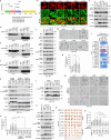
a The MOB1A/B LOF missense mutants fail to rescue YAP cytoplasmic localization in the MOB1A/B DKO cells. Scale bar, 30 μm. Arrows showed the cells expressing the indicated constructs. b, c The MOB1A/B LOF mutations inhibit MOB1A/B functions. The MOB1A/B DKO HEK293A cells were transduced with the indicated constructs and subjected to Western blot (b). A representative blot of two independent experiments is shown. The transcription of YAP downstream genes CTGF, CYR61 and ANKRD1 was examined by q-PCR (mean ± s.d., n = 3 biological replicates) (c). **p < 0.01, ***p < 0.001 (two-tailed Student’s t-test). d The MOB1A/B LOF mutants fail to rescue YAP S127 phosphorylation in the MOB1A/B DKO CAL-27 cells. A representative blot of two independent experiments is shown. e, f The MOB1A/B LOF mutants fail to inhibit MOB1A/B-deficient CAL-27 xenograft tumor growth in mouse tongues. The collected mouse tongues were shown (e). Tumor volume was measured and quantified (mean ± s.d., n = 6 mice per group) (f). ns, no significance. ***p < 0.001 (two-tailed Student’s t-test). g, h The MOB1A/B LOF missense mutations inhibit their interaction with LATS1. A representative blot of two independent experiments is shown. i The MOB1A/B LOF missense mutations inhibit their phosphorylation at T12 and T35. A representative blot of two independent experiments is shown. j The MOB1A/B LOF missense mutations inhibit MST1-mediated their T35 phosphorylation in vitro. A representative blot of two independent experiments is shown. k, l The MOB1A/B protein structures are changed by their LOF mutations. m Illustration of the MOB1 ZNF domain in the p-MOB1/LATS1-MBD co-crystal structure (PDB: 5BRK). np The MOB1A ZNF domain mutations target MOB1A function in the Hippo pathway. The MOB1A/B DKO HEK293A cells were transfected with the indicated constructs and subjected to immunofluorescent staining (n) and Western blot (o). Scale bar, 30 μm. Arrows showed the cells expressing the indicated constructs. HEK293T cells were transfected with Myc-LATS1 and the indicated SFB-MOB1A/B constructs and subjected to pulldown assay (p). A representative blot of two independent experiments is shown. q The MOB1A protein structure is changed by its ZNF domain site mutations. r MOB1A protein structure is changed by TPEN. MBP-tagged MOB1A protein was purified from bacteria, incubated with TPEN at the indicated concentrations, and subjected to circular dichroism analysis. s TPEN treatment disrupts the MOB1-LATS1 complex formation. Cell lysates were incubated with TPEN at different concentrations (i.e., 0 mM, 1 mM, 2 mM, and 4 mM) and subjected to pulldown assay. A representative blot of two independent experiments is shown. Source data are provided as a Source Data file.
Fig. 7
Fig. 7. The NF2 LOF mutations induce an oncogenic role of NF2 through the VANGL-JNK pathway.
a Illustration of NF2 domains and its identified LOF missense mutations. b The NF2 LOF mutants fail to rescue YAP cytoplasmic localization in the NF2 KO cells. Scale bar, 30 μm. Arrows showed the cells expressing the indicated constructs. c Reconstituting the NF2 LOF mutants fail to rescue YAP S127 phosphorylation in the NF2 KO cells. A representative blot of two independent experiments is shown. d, e The NF2 LOF mutations inhibit its interaction with LATS1 and AMOT. HEK293T cells were transfected with the indicated HA-NF2 constructs and SFB-tagged LATS1 (d) or AMOT (e) and subjected to pulldown assay. A representative blot of two independent experiments is shown. f NF2 LOF mutants induces the phosphorylation of JNK and c-Jun in MCF10A cells. A representative blot of two independent experiments is shown. g, h The NF2 LOF mutants promote MCF10A acini growth. The representative images were shown (g). The acini size was measured and quantified (mean ± s.d., n = 40 acini per group) (h). ***p < 0.001 (two-tailed Student’s t-test). Scale bar, 400 μm. i, j The NF2 LOF mutations enhance its interaction with VANGL1. The interaction between the indicated HCIPs and NF2 LOF mutants was normalized and shown as a heatmap (i). HEK293T cells were transfected with SFB-VANGL1 and the indicated HA-NF2 LOF mutants and subjected to pulldown assay (j). A representative blot of two independent experiments is shown. k The NF2 LOF mutants activate JNK through VANGL1. The MCF10A cells stably expressing NF2 and its LOF mutants were transduced with control and VANGL1 shRNAs. A representative blot of two independent experiments is shown. l The MCF10A cells stably expressing the NF2 LOF mutants were treated with JNK-IN-8 (10 μM) for 4 h and subjected to Western blot. A representative blot of two independent experiments is shown. m, n VANGL1 and JNK are required for the NF2 LOF mutants-induced MCF10A acini growth in Matrigel. The representative images were shown (m). The acini size was measured and quantified (mean ± s.d., n = 40 acini per group) (n). ***p < 0.001 (two-tailed Student’s t-test). Scale bar, 400 μm. o The NF2 LOF mutants activate JNK in IOMM-Lee cells. A representative blot of two independent experiments is shown. p, q Treatment with JNK-IN-8 inhibits NF2 LOF mutants-induced IOMM-Lee xenograft tumor growth. The collected tumors treated with DMSO or JNK-IN-8 (10 mg/kg) were shown (p) and tumor weight was measured and quantified (mean ± s.d., n = 6 mice per group) (q). ns, no significance. *p < 0.05, **p < 0.01, ***p < 0.001 (two-tailed Student’s t-test). Source data are provided as a Source Data file.

Similar articles

Cited by

References

    1. Yu, F. X., Zhao, B. & Guan, K. L. Hippo pathway in organ size control, tissue homeostasis, and cancer. Cell163, 811–828 (2015). - PMC - PubMed
    1. Ma, S., Meng, Z., Chen, R. & Guan, K. L. The Hippo pathway: biology and pathophysiology. Annu Rev. Biochem88, 577–604 (2019). - PubMed
    1. Zheng, Y. & Pan, D. The Hippo signaling pathway in development and disease. Dev. Cell50, 264–282 (2019). - PMC - PubMed
    1. Halder, G. & Johnson, R. L. Hippo signaling: growth control and beyond. Development138, 9–22 (2011). - PMC - PubMed
    1. Yin, F. et al. Spatial organization of Hippo signaling at the plasma membrane mediated by the tumor suppressor Merlin/NF2. Cell154, 1342–1355 (2013). - PMC - PubMed

Publication types

MeSH terms

Substances