

OCTA evaluates changes in retinal microvasculature in renal hypertension patients
- PMID: 39572632
- PMCID: PMC11582710
- DOI: 10.1038/s41598-024-68690-3
OCTA evaluates changes in retinal microvasculature in renal hypertension patients
Abstract
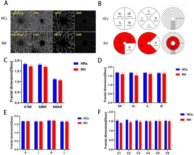
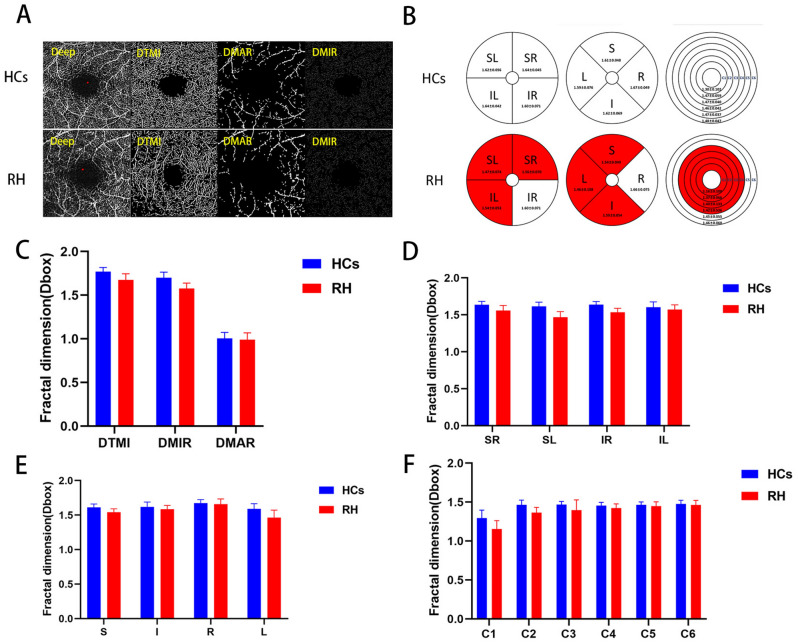
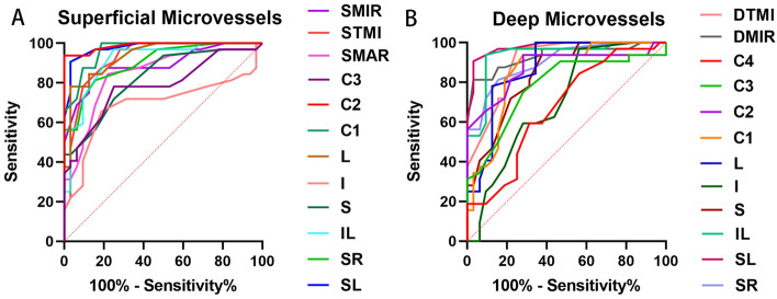
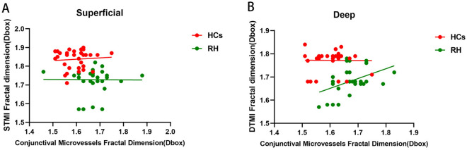
The objective of this study is to utilize optical coherence tomography angiography (OCTA) techniques for the purpose of identifying abnormalities in retinal and conjunctival vascular density among patients afflicted with renal hypertension. From October 2022 to October 2023, a cohort of sixteen patients diagnosed with renal hypertension (RH), comprising a total of 32 eyes, was selected from the Department of Nephrology at the First Affiliated Hospital of Nanchang University. Concurrently, a group of sixteen healthy individuals, carefully matched in characteristics, was recruited from volunteers at the Ophthalmology Research Center and designated as the healthy controls (HCs) group. Optical coherence tomography angiography was employed to assess and examine the superficial vascular plexus (SVP) and deep vascular plexus (DVP) of the macular retina in both eyes. Subsequently, a comparative analysis was conducted between the two groups, focusing on the superficial and deep retinal microvessels (MIR), macrovascular (MAR), and total microvascular (TMI). The present study employed the central annuli segmentation method (C1-C6), the hemispheric segmentation method (SL, IL, SR, IR), and the Early Treatment Diabetic Retinopathy Study (S, I, L, R) to evaluate deviations in retinal blood vessel density. The investigation aimed to examine the association between blood vessel density and TMI in conjunctival capillaries. A statistically significant difference (p < 0.05) in macular retinal vascular density was observed between the two groups based on the OCTA data. Specifically, in SVP, the density of TMI, MIR, and MAR in the RH group was significantly lower compared to the HCs group (p < 0.05). Additionally, the deep density of TMI and MIR in DVP of the RH group was significantly lower than that of the HCs group (p < 0.05). Furthermore, using the hemispheric segmentation method, both the superficial and deep retina showed a significant reduction in the density of SL, SR and IL regions (p < 0.05). In the ETDRS method, there was a significant decrease in superficial and deep retinal S, I, and L in the RH group (p < 0.05). When applying the central annuli segmentation methods, the RH group exhibited a significant decrease in the superficial retinal C1-3 region (p < 0.05) and a noticeable reduction in the deep retina in the C1-4 region (p < 0.05). Furthermore, a higher positive likelihood ratio was observed in the deep SL and superficial C2 region. There was a positive correlation between conjunctival capillary density and the region of TMI in depth. The results of the OCTA investigation revealed a significant disparity in the density of superficial and deep retinal blood vessels between RH group and the HCs group. Additionally, a notable correlation was observed between the depth of TMI and the density of conjunctival capillaries. These findings highlight the potential of retinal OCTA as a valuable tool for early detection and image-assisted diagnosis of retinopathy progression in patients with RH.
Keywords: Microvessel density; Optical coherence tomography angiography; Renal hypertension.
© 2024. The Author(s).
Conflict of interest statement
Competing interests: The authors declare no competing interests.
Figures

References
-
- Ma, Y. J., Dong, Y. X. & He, Y. Diagnosis and treatment of renal hypertension in clinic. Med. Innov. China21(01), 179–184 (2024).
-
- Whelton, P. K. et al. ACC/AHA/AAPA/ABC/ACPM/AGS/APhA/ASH/ASPC/NMA/PCNA guideline for the prevention, detection, evaluation, and management of high blood pressure in adults: Executive summary: A report of the American college of cardiology/American heart association task force on clinical practice guidelines. Hypertension71(6), 1269–1324 (2018). - DOI - PubMed
-
- Zhai, R. N., Zheng, L. Y., Xue, R., Gui, D. K. & Wang, N. S. Research progress on renal hypertension pathogenesis and its diagnosis and treatment. World Clin. Drug38(5), 305–310 (2017).
MeSH terms
Grants and funding
- No.82160195/National Natural Science Foundation of China
- No.jxsq2023201036/Jiangxi Double-Thousand Plan High-Level Talent Project of Science and Technology Innovation
- 20223BBH80014/Key R & D Program of Jiangxi Province
- No. 2022B258/Science and Technology Project of Jiangxi Province Health Commission of Traditional Chinese Medicine
- No. 202210017/Science and Technology Project of Jiangxi Health Commission
LinkOut - more resources
Full Text Sources
Research Materials
Miscellaneous
