. 2024 Nov 21;7(1):1554.
doi: 10.1038/s42003-024-07242-x.

Plasmodium berghei liver stage parasites exploit host GABARAP proteins for TFEB activation

Affiliations

Plasmodium berghei liver stage parasites exploit host GABARAP proteins for TFEB activation

Jacqueline Schmuckli-Maurer et al. Commun Biol. .

Abstract

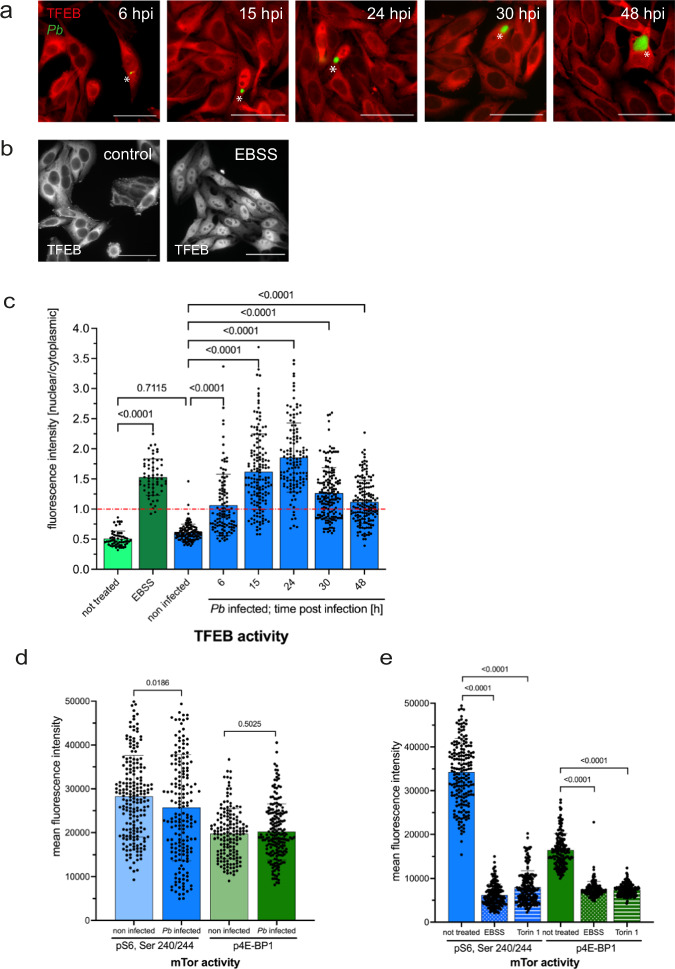
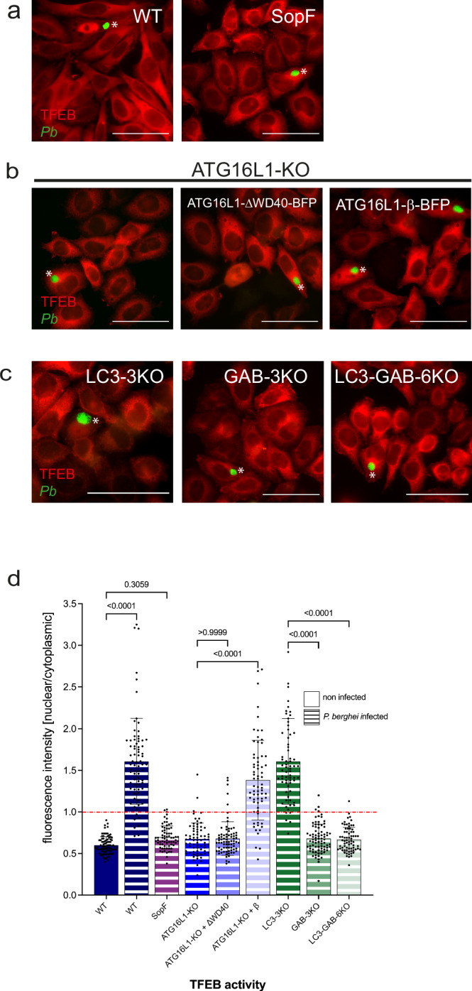
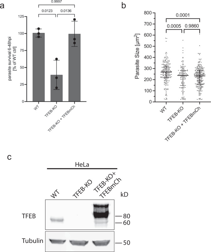
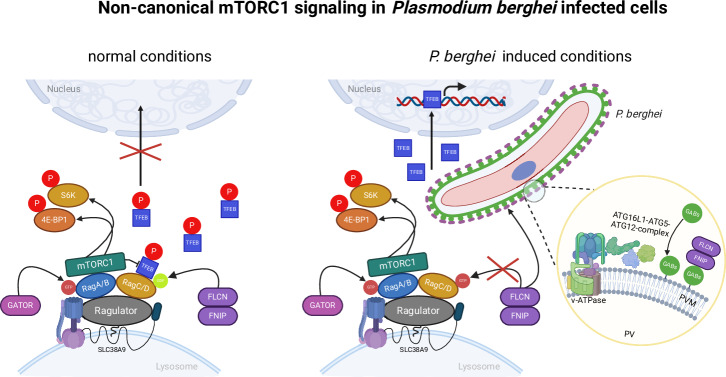
Plasmodium, the causative agent of malaria, infects hepatocytes prior to establishing a symptomatic blood stage infection. During this liver stage development, parasites reside in a parasitophorous vacuole (PV), whose membrane acts as the critical interface between the parasite and the host cell. It is well-established that host cell autophagy-related processes significantly impact the development of Plasmodium liver stages. Expression of genes related to autophagy and lysosomal biogenesis is orchestrated by transcription factor EB (TFEB). In this study, we explored the activation of host cell TFEB in Plasmodium berghei-infected cells during the liver stage of the parasite. Our results unveiled a critical role of proteins belonging to the Gamma-aminobutyric acid receptor-associated protein subfamily (GABARAP) of ATG8 proteins (GABARAP/L1/L2 and LC3A/B/C) in recruiting the TFEB-blocking FLCN-FNIP (Folliculin-Folliculin-interacting protein) complex to the PVM. Remarkably, the sequestration of FLCN-FNIP resulted in a robust activation of TFEB, reliant on conjugation of ATG8 proteins to single membranes (CASM) and GABARAP proteins. Our findings provide novel mechanistic insights into host cell signaling occurring at the PVM, shedding light on the complex interplay between Plasmodium parasites and the host cell during the liver stage of infection.


Conflict of interest statement

Competing interests: The authors declare the following competing interests: L.O.M. and T.L. are employees and shareholders of Casma Therapeutics. A.B. is a co-founder and shareholder of CASMA Therapeutics, Inc, and Advisory board member of Avilar Therapeutics and Amplify Therapeutics. L.O.M. is an author on a patent related to TFEB modulation, in addition. M.L. is a co-founder and member of the scientific advisory board of Automera. Other authors declare no competing interests.

Figures

Fig. 1
Fig. 1. TFEB translocates to the host cell nucleus upon P. berghei infection.
a HeLa cells constitutively expressing TFEBmCh were infected with PbGFP sporozoites. Samples were fixed at 6, 15, 24, 30, and 48 hpi and stained with anti-GFP (green) and anti-RFP (red) antibodies to enhance the signals. Samples were analyzed with a widefield fluorescent microscope. Parasites are labeled with a white asterisk. Scale bar 50 µm. Note that in infected cells, TFEBmCh locates to the host cell nucleus whereas in non-infected cells TFEBmCh is in the cytoplasm. b HeLa cells expressing TFEBmCh were grown in full medium (control) or in starvation medium (EBSS) for 2 h. Samples were fixed and stained with anti-RFP antibodies to enhance the TFEB signal here shown in gray. Only in starved cells TFEB localizes to the cell nucleus. c Quantification of the experiment described in (a) and (b). Cells were fixed at indicated time points and stained with anti-GFP (only infected cells) and anti-RFP antibodies, pictures were taken with a widefield fluorescent microscope. Fluorescence intensity in the nucleus and the cell cytoplasm was measured and the ratio nuclear/cytoplasmic was calculated for each cell. A ratio above 1 indicates more nuclear than cytoplasmic TFEB, a ratio lower than 1 indicates more cytoplasmic than nuclear TFEB. Note that starved and Pb-infected cells have a ratio above 1 which means activated TFEB. Pictures were analyzed using Fiji. N > 30 for each cell line in each experiment. The graph depicts mean and SD of the pooled data of two independent experiments. P-values were calculated using a one-way ANOVA test. d mTORC1 activity in Pb-infected HeLa cells. Non-infected and PbmCh-infected cells were fixed 24 hpi and stained with anti-pS6(Ser240/244) or with anti-p4E-BP1 antibodies to visualize phosphorylated substrates of the mTOR kinase. Note that mTOR is active in Pb-infected cells at the same level as in non-infected cells. Pictures were taken with a widefield fluorescent microscope and fluorescence intensity was measured using Fiji. N > 60 for each sample in each experiment. The graph depicts mean and SD of the pooled data of two independent experiments. P-values were calculated using a Student’s t test. e Control experiment for experiment described in (d). mTORC1 activity in non-treated, starved (EBSS, 2 h) and Torin 1 (200 nM, 2 h) treated HeLa cells. Cells were fixed and stained with anti-pS6(Ser240/244) or with anti-p4E-BP1 antibodies to visualize phosphorylated substrates of the mTOR kinase. Note that Torin 1 and EBSS inhibit mTOR kinase. Experiment was performed as described in (d).
Fig. 2
Fig. 2. P. berghei induced TFEB nuclear translocation depends on CASM and GABARAP proteins.
a SopF inhibits TFEB nuclear translocation in Pb-infected HeLa cells. TFEBmCh, SopF expressing HeLa cells were infected with PbGFP sporozoites. 24 hpi cells were fixed and stained with anti-GFP (green) and anti-RFP (red) antibodies to enhance the signals. Samples were analyzed with a widefield fluorescence microscope. Parasites are labeled with a white asterisk. Scale bar 50 µm. Note, in Pb-infected WT cells TFEB always localizes to the host cell nucleus, whereas in SopF expressing cells TFEB is cytoplasmic, even in infected cells. b Pb induced TFEB nuclear translocation depends on ATG16L1-WD40 domain. HeLa cells lacking ATG16L1, reconstituted with either ATG16L1-ΔWD40-BFP or with ATG16L1-β-BFP all expressing TFEBmCh, were infected with PbGFP sporozoites. 24 hpi cells were treated as described in (a). Parasites are labeled with a white asterisk. Scale bar 50 µm. Note that nuclear TFEB can be found only in ATG16L1-KO cells expressing the WT ATG16L1-β-BFP. In ATG16L1-KO cells or KO cells reconstituted with ATG16L1-ΔWD40-BFP TFEB is trapped in the cytoplasm. c GABARAPs are indispensable for Pb induced TFEB nuclear translocation. TFEBmCh expressing HeLa cells lacking all 3 LC3s (LC3A, LC3B, LC3C), or all 3 GABARAPs (GAB, GABL1, GABL2) or lacking all LC3s and all GABs were infected with PbGFP. 24 hpi cells were treated as described in (a). Parasites are labeled with a white asterisk. Scale bar 50 µm. Note that TFEB nuclear translocation only happens in the cell line expressing GABARAPs. d Quantification of the experiments described in (a), (b), and (c). All cell lines constitutively express TFEBmCh and were infected with PbGFP. Cells were fixed 24 hpi and stained with anti-GFP (only infected cells) and anti-RFP antibodies, pictures were taken with a widefield fluorescence microscope. Fluorescence intensity in the nucleus and the cell cytoplasm was measured and the ratio nuclear/cytoplasmic was calculated for each cell A ratio above 1 indicates more nuclear than cytoplasmic TFEB, a ratio lower than 1 indicates more cytoplasmic than nuclear TFEB. Pictures were analyzed using Fiji. N > 30 for each cell line in each experiment. The graph depicts mean and SD of the pooled data of two independent experiments. P-values were calculated using a one-way ANOVA test. Note that Pb induced TFEB nuclear translocation depends on the ATG16L1-WD40 domain and on GABARAPs and can be inhibited by SopF.
Fig. 3
Fig. 3. GABARAPs localize to Plasmodium PVM depending on ATG16L1.
a All 3 GABARAP proteins localize to Pb PVM. HeLa WT cells were transiently transfected with GFP-GABARAPs and approximately 15 h post transfection infected with PbmCh sporozoites. 6 hpi infected cells were fixed and stained with anti-GFP antibodies (green) to enhance the GABARAP signal and anti-UIS4 antibodies (magenta) to visualize the Pb PVM. DNA was stained with Dapi (cyan). Images were taken with a confocal laser scanning microscope. Scale bar 5 µm. Note that GABARAP, GABARAPL1, and GABARAPL2 clearly localize to the P. berghei PVM. b Quantification of (a). Graph shows the Pearson’s correlation coefficient (PCC) for UIS4 and GFP-GABARAPs. PCC was calculated using the Coloc2 tool of FIJI. N = 5 parasites. Each dot represents one parasite, each red dot represents the parasites shown in (a). c PVM localization of GABARAPs depends on ATG16L1. HeLa cells lacking ATG16L1 were transiently transfected with GFP-GABARAPs and treated as described in (a). Images were taken with a confocal laser scanning microscope. Scale bar 5 µm. Note that none of the GABARAP proteins localizes to the Pb PVM in ATG16L1-KO cells. d Quantification of the experiments described in (a) and (c). The graph shows the percentage of GABARAP-positive parasites in HeLa WT and ATG16L1-KO cells. Only UIS4-positive parasites were counted. The graph depicts the mean and SD of two independent experiments. P-values were calculated using a Student’s t test. N 70 per experiment and cell line. e GABARAPs are needed for nuclear translocation of TFEB in Pb-infected cells. Quantification of the TFEB signal in GAB-3KO cells and GAB-3KO cells transiently transfected with each of the GFP-GABARAPs all constitutively expressing TFEBmCh. Cells were fixed 24 hpi and stained with anti-GFP and anti-RFP antibodies, pictures were taken with a widefield fluorescence microscope. Fluorescence intensity in the nucleus and the cell cytoplasm was measured and the ratio nuclear/cytoplasmic was calculated for each cell. A ratio above 1 indicates more nuclear than cytoplasmic TFEB, a ratio lower than 1 indicates more cytoplasmic than nuclear TFEB. Note that all GABARAPs are proficient to activate TFEB upon Pb infection. Pictures were analyzed using Fiji. N > 30 for each cell line in each experiment. The graph depicts mean and SD of the pooled data of two independent experiments. P-values were calculated using a one-way ANOVA test.
Fig. 4
Fig. 4. FNIP1 localizes to Pb PVM depending on GABARAPs and on FNIP1-LIR domain.
a FNIP1 localization at P. berghei PVM. HeLa WT, GAB3-KO, and FNIP1/2-DKO cells all constitutively expressing 3x-HA-FNIP1 were infected with PbmCh. 6 hpi cells were fixed and stained with anti-HA (green) and anti-UIS4 (magenta). PbmCh is shown in red. DNA was stained with Dapi (cyan). Images were taken with a confocal laser scanning microscope. Scale bar 10 µm. Note that the PVM localization of FNIP1 is dependent on GABARAPs and on a functional FNIP1-LIR domain. b Quantification of the experiment described in (a). The graph shows the percentage of FNIP1-positive parasites in the different HeLa cell lines. Only UIS4-positive parasites were counted. The graph depicts the mean and SD of two independent experiments. P-values were calculated using a Student’s t test. N 70 per experiment and cell line. c Quantification of the experiment described in (a). Graph shows the Pearson’s correlation coefficient (PCC) for UIS4 and 3xHA-FNIP1. PCC was calculated using the Coloc2 tool of FIJI. N = 11 parasites for HeLa WT, 7 parasites for GAB-3KO, 6 for FNIP1/2-DKO + 3xHA-FNIP1, and 6 parasites for FNIP1/2-DKO + 3xHA-FNIP1-LIR. Each symbol represents one parasite, each red symbol represents the parasites shown in (a). d TFEB activation in infected cells depends on Pb PVM localization of FNIP1. All cell lines constitutively express TFEBmCh. FNIP1-DKO cells additionally express 3xHA-FNIP1, 3xHA-FNIP1-LIR or are transiently transfected with HA-RagD77L (a constitutively active mutant of RagD), as indicated. Cells were infected with PbmCh and fixed 24 hpi and stained with anti-RFP and anti-HA (only RagD transfected). Pictures were taken with a widefield fluorescence microscope. Fluorescence intensity in the nucleus and the cell cytoplasm was measured and the ratio nuclear/cytoplasmic was calculated for each infected cell. A ratio above 1 indicates that more TFEB is in the nucleus than in the cytoplasm and a ratio lower than 1 indicates mor TFEB in the cytoplasm than in the nucleus. Pictures were analyzed using Fiji. N > 50 for each cell line in each experiment. The graph depicts mean and SD of the pooled data of two independent experiments. Single-color bars represent non-infected cells, striped bars represent Plasmodium-infected cells. P-values were calculated using a one-way ANOVA test. Note that TFEB is only activated when FNIP1 is sequestered to the Pb PVM. e Starvation control for the cell lines used in the experiment described in (d). Cells were left untreated (single-color bars) or starved for 2 h in EBSS (dotted bars). The analysis was carried out as described in (d). N > 50 for each cell line in each experiment. The graph depicts mean and SD of the pooled data of two independent experiments. P-values were calculated using a one-way ANOVA test. Note that a functional FNIP1 LIR domain is not needed for TFEB activation in starved cells.
Fig. 5
Fig. 5. TFEB supports parasite survival.
a P. berghei survival is reduced in TFEB-KO cells. HeLa WT, TFEB-KO, and TFEB-KO + TFEBmCh cells were infected with PbGFP. At 6 and 48 hpi, parasite numbers were evaluated using automated high throughput live cell imaging and analysis (INCell Analyzer 2000). The graph shows relative parasite survival from 6 to 48 hpi compared to the WT control. The mean and SD of three independent experiments are depicted. N > 500 parasites per cell line and experiment. A one-way ANOVA test was used to determine p-values. b Parasite size at 48 hpi of the experiment described in (a). The experiment was performed three times. Shown is the data of one representative experiment. Median and SD are depicted. P-values were calculated using a one-way ANOVA test. HeLa WT N = 193; TFEB-KO N = 129; TFEB-KO + TFEBmCh N = 237. c Western blot of the cell lines used in (a) and (b). Whole protein lysates were separated on a 12% acrylamide gel, transferred onto a nitrocellulose membrane, and probed with an anti-TFEB antibody. α-tubulin was detected as a loading control. Note that in line 3, the TFEBmCh fusion protein expressed in the TFEB-KO cells is larger than the endogenously expressed TFEB due to the mCherry fusion partner (see also Fig. S8a, b).
Fig. 6
Fig. 6. Schematic overview of transcription factor EB activation in Plasmodium berghei-infected cells.
Normal conditions: active mTORC1 is localized to lysosomes by binding to RagA/B, which in turn binds to the Ragulator complex. This complex interacts with the V-ATPase located in the lysosomal membrane. The FLCN-FNIP1/2 complex, acting as a GTPase-activating protein (GAP) for RagC/D, enables active RagC/D (bound to GDP) to directly interact with TFEB. This interaction positions TFEB near mTORC1, leading to TFEB phosphorylation. Phosphorylated TFEB is then sequestered in the cytoplasm by its binding to a 14-3-3 protein. Active mTORC1 also phosphorylates numerous other substrates, such as 4E-BP1 and S6K, promoting anabolic processes like protein, nucleotide, and lipid synthesis while inhibiting catabolic processes like autophagy and lysosome biogenesis. P. berghei-induced TFEB activation: host cell GABARAP proteins are integrated into the parasitophorous vacuolar membrane (PVM) of the parasite shortly after infection, via a mechanism involving V-ATPase-ATG16L1-ATG5-ATG12 (CASM). This leads to the sequestration of the FLCN/FNIP complex at the PVM through interactions with GABARAPs, disrupting its GAP activity. Consequently, RagC/D becomes inactive (bound to GTP), preventing its interaction with TFEB. Without this interaction, TFEB remains unphosphorylated and moves into the nucleus to activate target genes. Meanwhile, mTORC1 remains active and bound to RagA/B, capable of phosphorylating its substrates. In cells infected by P. berghei, we observe a unique scenario where both anabolic and catabolic processes are simultaneously activated within the same cell. Schematic created with BioRender.com.

References

    1. Frischknecht, F. et al. Imaging movement of malaria parasites during transmission by Anopheles mosquitoes. Cell. Microbiol.6, 687–694 (2004). - PubMed
    1. Amino, R. et al. Quantitative imaging of Plasmodium transmission from mosquito to mammal. Nat. Med.12, 220–224 (2006). - PubMed
    1. Tavares, J. et al. Role of host cell traversal by the malaria sporozoite during liver infection. J. Exp. Med.210, 905–915 (2013). - PMC - PubMed
    1. Pradel, G. & Frevert, U. Malaria sporozoites actively enter and pass through rat Kupffer cells prior to hepatocyte invasion. Hepatology33, 1154–1165 (2001). - PubMed
    1. Sturm, A. et al. Manipulation of host hepatocytes by the malaria parasite for delivery into liver sinusoids. Science313, 1287–1290 (2006). - PubMed

MeSH terms

Substances