. 2024 Nov 21;14(1):28913.
doi: 10.1038/s41598-024-80411-4.

Suppression of renal crystal formation, inflammation, and fibrosis by blocking oncostatin M receptor β signaling

Affiliations

Suppression of renal crystal formation, inflammation, and fibrosis by blocking oncostatin M receptor β signaling

Ryusuke Deguchi et al. Sci Rep. .

Abstract

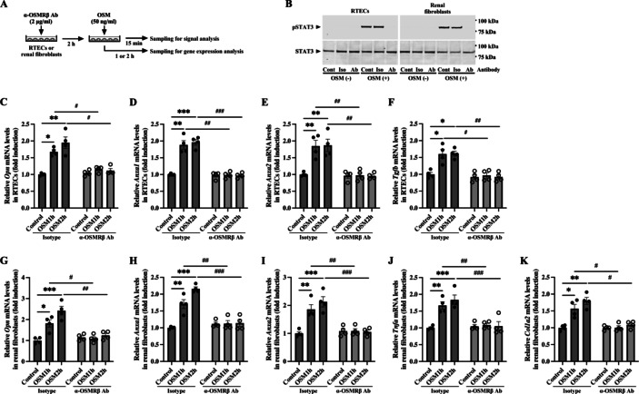
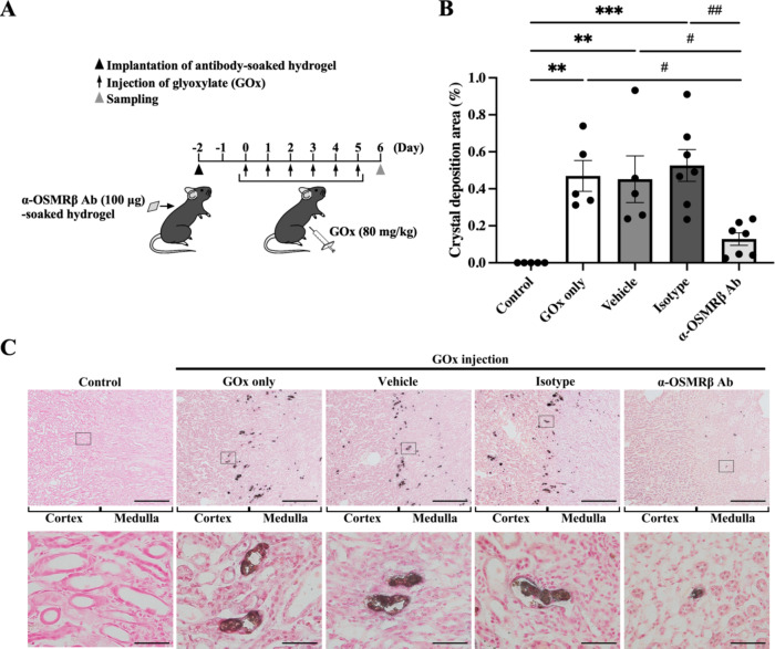
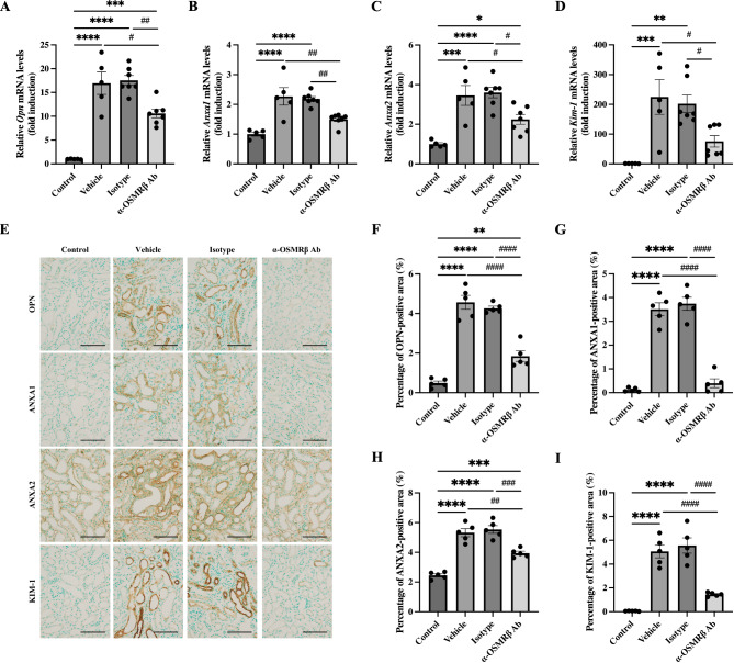
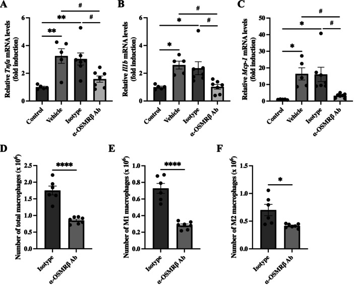
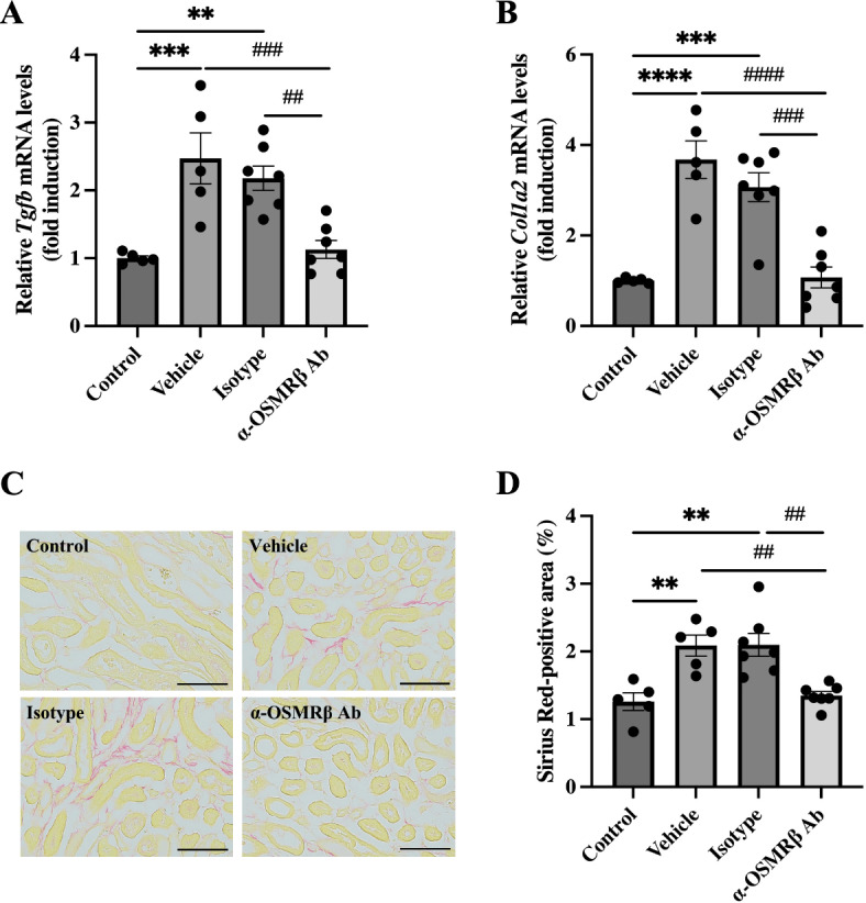
Oncostatin M (OSM) has pleiotropic effects on various inflammatory diseases, including kidney stone disease. The prevalence of kidney stones has increased worldwide, despite recent therapeutic advances, due to its high recurrence rate, suggesting the importance of prevention of repeated recurrence in the treatment of kidney stone disease. Using a mouse model of renal crystal formation, we investigated the preventive effects of blockade of OSM receptor β (OSMRβ) signaling on the development of kidney stone disease by treatment with a monoclonal anti-OSMRβ antibody that we generated. The anti-OSMRβ antibody abrogated OSM-induced phosphorylation of STAT3 and expression of crystal-binding molecules (Opn, Anxa1, Anxa2) and inflammation/fibrosis-associated molecules (Tnfa, Tgfb, Col1a2) in renal tubular epithelial cells and fibroblasts. In glyoxylate-injected mice, a mouse model of renal crystal formation, there was significant suppression of crystal deposits and expression of crystal-binding molecules (Opn, Anxa1, Anxa2), a tubular injury marker (Kim-1), and inflammation/fibrosis-associated molecules (Tnfa, Il1b, Mcp-1, Tgfb, Col1a2) in the kidneys of the anti-OSMRβ antibody-treated mice, compared with those in vehicle- or isotype control antibody-treated mice. In addition, treatment with the anti-OSMRβ antibody significantly decreased infiltrating macrophages and fibrosis in the kidneys. These findings suggest that anti-OSMRβ antibody-treatment may be effective in preventing kidney stone disease.


Conflict of interest statement

Declarations. Competing interests: T. Komori and Y. Morikawa are the inventors on a patent for the use of anti-OSMRβ antibody (7D2) in treatment of atopic dermatitis (US9475876B2; JP6214010). T. Komori, S. Yamashita, Y. Kohjimoto, I. Hara, and Y. Morikawa have a patent pending on the use of anti-OSMRβ antibody (7D2) in treatment of kidney stone disease. All other authors declare they have no conflict of interest.

Figures

Fig. 1
Fig. 1
The anti-OSMRβ antibody inhibited the effects of OSM on the RTECs and renal fibroblasts. (A) Experimental design for in vitro effects of the anti-OSMRβ antibody on OSM-induced responses. (B) Suppression of OSM-induced STAT3 activation by the anti-OSMRβ antibody. Western blot analysis of pSTAT3 (upper panels) and STAT3 (lower panels) in the RTECs (left panels) and renal fibroblasts (right panels). The positions of molecular weight markers are indicated on the right of the panels. The full-length blots are presented in Supplementary Fig. 3. Cont cells treated with vehicle, Iso cells treated with the isotype control antibody, Ab cells treated with the anti-OSMRβ antibody. (C–K) Effects of the anti-OSMRβ antibody on the expressions of genes induced by OSM in the RTECs (C–F) and renal fibroblasts (G–K) (n = 4). Data are expressed as mean ± SEM. *p < 0.05, **p < 0.01, ***p < 0.001 (versus Control); #p < 0.05, ##p < 0.01, ###p < 0.001 (versus Isotype). Two-way ANOVA followed by Tukey’s post hoc test.
Fig. 2
Fig. 2
The anti-OSMRβ antibody suppressed formation of renal crystal deposits in a mouse model of renal crystal formation. (A) Experimental design for in vivo effects of the anti-OSMRβ antibody on GOx-induced mouse model of renal crystal formation. (B) Quantification of renal crystal deposits by Pizzolato staining. The renal crystal deposits were quantified as the percentage of the area containing crystal deposits to the total kidney area (n = 5–7 per group). (C) Representative images of Pizzolato staining in the kidneys of mice on day 6. The boxed areas in the upper panels are shown at higher magnification in the corresponding lower panels. The sections were counterstained with nuclear fast red. Scale bars = 500 μm (upper panels); 50 μm (lower panels). Data are expressed as mean ± SEM. **p < 0.01, ***p < 0.001 (versus Control); #p < 0.05, ##p < 0.01 (versus α-OSMRβ Ab). Two-way ANOVA followed by Tukey’s post hoc test.
Fig. 3
Fig. 3
Suppressive effects of the anti-OSMRβ antibody on the expression of crystal-binding protein in the kidneys of a mouse model of renal crystal formation. (A–D) Gene expressions of Opn (A), Anxa1 (B), Anxa2 (C), and Kim-1 (D) in the kidneys of mice on day 6 (n = 5–7 per group). (E) Representative images of immunohistochemical staining for OPN, ANXA1, ANXA2 and KIM-1 in the kidneys of mice on day 6. The sections were counterstained with methyl green. Scale bars = 100 μm. (F–I) Quantification of immunohistochemical staining for OPN (F), ANXA1 (G), ANXA2 (H) and KIM-1 (I) (n = 5 per group). Data are expressed as mean ± SEM. *p < 0.05, **p < 0.01, ***p < 0.001, ****p < 0.0001 (versus Control); #p < 0.05, ##p < 0.01, ###p < 0.001, ####p < 0.0001 (versus α-OSMRβ Ab). Two-way ANOVA followed by Tukey’s post hoc test.
Fig. 4
Fig. 4
Suppressive effects of the anti-OSMRβ antibody on renal inflammation in the kidneys of a mouse model of renal crystal formation. (A–C) Gene expressions of Tnfa (A), Il1b (B), and Mcp-1 (C) in the kidneys of mice on day 6 (n = 5–7 per group). (D–F) Flowcytometric analysis of total macrophages (D), M1 macrophages (E), and M2 macrophages (F) in the kidneys of mice on day 6 (n = 6–7 per group). Data are expressed as mean ± SEM. *p < 0.05, **p < 0.01, ****p < 0.0001 (versus Control in A–C; versus Isotype in D–F); #p < 0.05 (versus α-OSMRβ Ab). Two-way ANOVA followed by Tukey’s post hoc test (A–C); Student’s t test (D–F).
Fig. 5
Fig. 5
Suppressive effects of the anti-OSMRβ antibody on fibrotic changes in the kidneys of a mouse model of renal crystal formation. (A,B) Gene expressions of Tgfb (A) and Col1a2 (B) in the kidneys of mice on day 6 (n = 5–7 per group). (C) Representative images of Sirius Red staining in the kidneys of mice on day 6. Scale bars = 100 μm. (D) Quantification of renal fibrosis by Sirius Red staining. The renal fibrosis was quantified as the percentage of the Sirius Red-positive area to the total kidney area (n = 5–7 per group). Data are expressed as mean ± SEM. **p < 0.01, ***p < 0.001, ****p < 0.0001 (versus Control); ##p < 0.01, ###p < 0.001, ####p < 0.0001 (versus α-OSMRβ Ab). Two-way ANOVA followed by Tukey’s post hoc test.

References

    1. Khan, S. R. et al. Kidney stones. Nat. Rev. Dis. Primers2, 16008 (2016). - PMC - PubMed
    1. Thongprayoon, C., Krambeck, A. E. & Rule, A. D. Determining the true burden of kidney stone disease. Nat. Rev. Nephrol.16, 736–746 (2020). - PubMed
    1. Rule, A. D. et al. The ROKS nomogram for predicting a second symptomatic stone episode. J. Am. Soc. Nephrol.25, 2878–2886 (2014). - PMC - PubMed
    1. Vaughan, L. E. et al. Predictors of symptomatic kidney stone recurrence after the first and subsequent episodes. Mayo Clin. Proc.94, 202–210 (2019). - PMC - PubMed
    1. Keddis, M. T. & Rule, A. D. Nephrolithiasis and loss of kidney function. Curr. Opin. Nephrol. Hypertens.22, 390–396 (2013). - PMC - PubMed

MeSH terms

Substances

LinkOut - more resources