. 2024 Nov 21;7(1):274.
doi: 10.1038/s42004-024-01344-7.

Preferential graphitic-nitrogen formation in pyridine-extended graphene nanoribbons

Affiliations

Preferential graphitic-nitrogen formation in pyridine-extended graphene nanoribbons

Nicolò Bassi et al. Commun Chem. .

Abstract

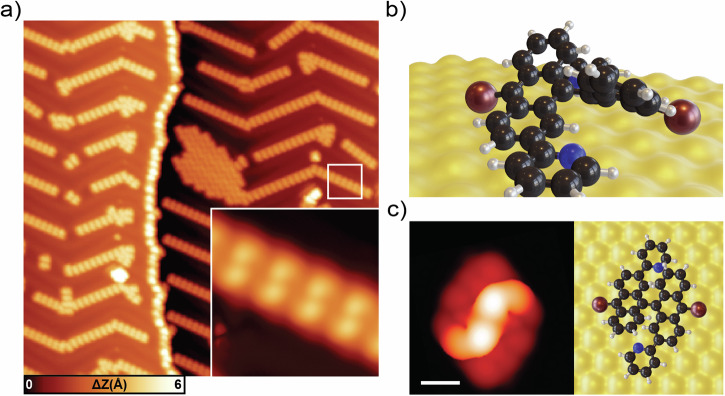
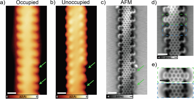
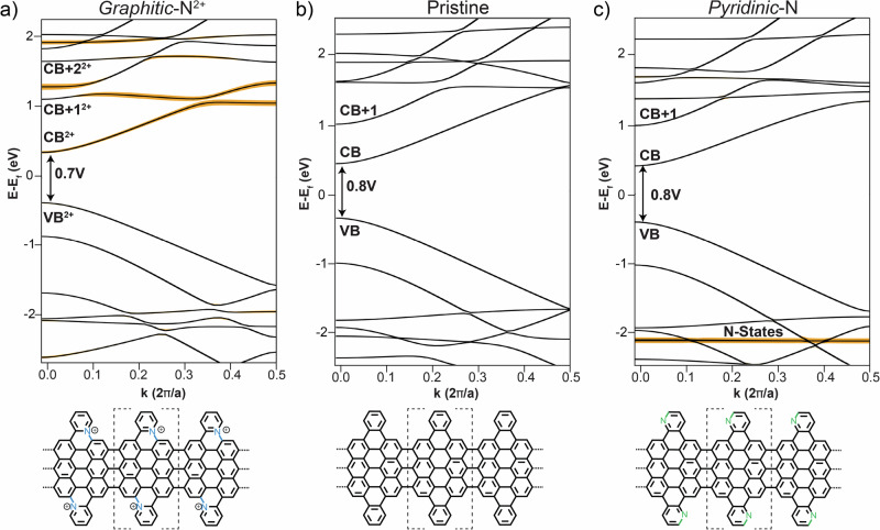
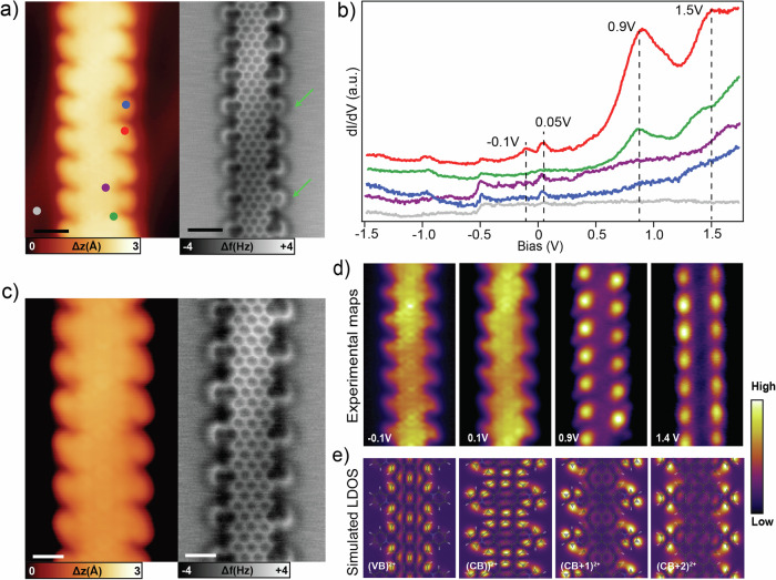
Graphene nanoribbons (GNRs), nanometer-wide strips of graphene, have garnered significant attention due to their tunable electronic and magnetic properties arising from quantum confinement. A promising approach to manipulate their electronic characteristics involves substituting carbon with heteroatoms, such as nitrogen, with different effects predicted depending on their position. In this study, we present the extension of the edges of 7-atom-wide armchair graphene nanoribbons (7-AGNRs) with pyridine rings, achieved on a Au(111) surface via on-surface synthesis. High-resolution structural characterization confirms the targeted structure, showcasing the predominant formation of carbon-nitrogen (C-N) bonds (over 90% of the units) during growth. This favored bond formation pathway is elucidated and confirmed through density functional theory (DFT) simulations. Furthermore, an analysis of the electronic properties reveals metallic behavior due to charge transfer to the Au(111) substrate accompanied by the presence of nitrogen-localized states. Our results underscore the successful formation of C-N bonds on the metal surface, providing insights for designing new GNRs that incorporate substitutional nitrogen atoms to precisely control their electronic properties.


Conflict of interest statement

Competing interests: The authors declare no competing interest.

Figures

Fig. 1
Fig. 1. Synthesis steps for Py-7-AGNRs.
Solution-synthesis of monomer 1 (upper row), used for the on-surface synthesis of Py-7-AGNRs (bottom row). The two possible configurations resulting in pyridinic-N and graphitic-N are highlighted in the last panel.
Fig. 2
Fig. 2. Self-assembly of DB-DPBA on Au(111).
a Large-scale STM topography after deposition of the molecular precursor. The molecules tend to organize in chains following the Au(111) herringbone reconstruction (scale bar: 20 nm, scanning parameters: −1 V, 50 pA). Inset: small-scale STM topography of the molecules organized in chains. The DB-DBPA are characterized by two brighter dots. b Equilibrium geometry of a single DB-DPBA obtained by DFT calculations (perspective view). The pyridyl units lay on the surface, while the bianthracene core is highly tilted. c DFT simulated STM image of a single molecule and top view of the equilibrium geometry (scale bar: 1 nm). There is good a matching with the STM appearance of molecules in the topography image in panel a.
Fig. 3
Fig. 3. Different annealing temperatures of DB-DBPA on Au(111).
a Large-scale STM topography after annealing at 180 °C. In the inset, a zoom-in image of a segment of a polymer with alternating dots with a period of 0.85 nm (scale bar: 5 nm, scanning parameters: −0.5 V, 30 pA). b Large-scale STM of partially planar segments after annealing at 250 °C. In the inset, a zoom-in image of partially planar segments (scale bar: 10 nm, scanning parameters: −0.5 V, 20 pA. At this temperature, some edges have already undergone planarization. In general, this process starts at the GNR end and proceeds along one edge. c Large-scale STM image of planar GNRs at 300 °C. In the inset, a zoom-in image of a segment of a ribbon (scale bar: 10 nm, scanning parameters: −0.3 V, 40 pA.) At this temperature, the segments are already completely planar.
Fig. 4
Fig. 4. Planar GNRs on Au(111) after annealing at 300 °C.
a STM topography image acquired in the occupied density of states. Most of the edge extensions have an upward (left side) or downward (right side) bend, with the exception of the two highlighted by green arrows (scale bar: 1 nm, scanning parameters:-1.0 V, 100 pA). b STM topography image of the same segment acquired in the unoccupied density of states. The edge extensions are imaged as bright dots, with the exception of the two indicated by green arrows (scale bar: 1 nm, scanning parameters: 0.80 V, 100 pA). c Bond-resolved nc-AFM image of the same segment of Py-7-AGNRs, revealing specific bonds in the pyridine extensions that appear darker. (open feedback parameters: −5 mV, 200 pA, Δz: 220 pm from Au(111) surface; frequency: 27401 Hz) (d) Nc-AFM image of a short segment of another Py-7-AGNR. The blue dashed box highlights a unit with fully graphitic-N edges, while the green box indicates a unit with pyridinic-N. (open feedback parameters: −5 mV, 200 pA, Δz: 210 pm from Au(111) surface; frequency: 27401 Hz) (e) DFT-based nc-AFM simulations of pyridinic-N (green) and graphitic-N (blue) units (scale bar: 1 nm).
Fig. 5
Fig. 5. Reaction barrier calculations for C-N and C-C bonds formation.
a Energy barrier for the C-N (blue) and C-C (green) bond formation as a function of atomic distances. In the inset, chemical sketch with dotted lines indicating the two controlled distances. The energy progressively increases as the distance is reduced until maxima are reached at 1.9 Å. After these points, the two curves decrease in energy thanks to the formation of bonds in the bianthracene backbone. b DFT-relaxed structures (perspective view) of different steps of the bond formations. In the right panel, the atoms involved are highlighted with red arrows.
Fig. 6
Fig. 6. Gas-phase DFT band structures of possible edge-extended 7-AGNRs.
Py-7-AGNR containing (a) graphitic-N1+ (b) pristine and (c) pyridinic-N. Bands with significant N contribution are highlighted by orange lines. The repetitive units are indicated by dotted square brackets in the chemical structure at the bottom of each panel. The graphitic-N1+ case is modeled with one electron per unit cell removed. For the band structure of the charge-neutral case, see Fig. SI 13c.
Fig. 7
Fig. 7. Electronic properties of Py-7-AGNRs.
a STM (left) and nc-AFM (right) image of a Py-7-AGNR with graphitic-N edges. Two pyridinic-N defects are labeled by green arrows. (scale bar: 1 nm, scanning parameters:−1.5 V, 100 pA). b dI/dV spectra on different points (see a). (Open feedback parameters: −1.5 V, 200 pA; Vrms: 20 mV) (c) STM (left) and nc-AFM (right) of another Py-7-AGNR segment without pyridinic-N defects. d dI/dV maps at selected bias voltages of the Py-7-AGNR shown in (c). e Simulated DOS maps of the singly charged system shown in Fig. 6a, evaluated at the different k-points of the corresponding bands.

References

    1. Cai, J. et al. Atomically precise bottom-up fabrication of graphene nanoribbons. Nature466, 470–473 (2010). - PubMed
    1. Talirz, L. et al. On-surface synthesis and characterization of 9-atom wide armchair graphene nanoribbons. ACS Nano11, 1380–1388 (2017). - PubMed
    1. Yamaguchi, J. et al. Small bandgap in atomically precise 17-atom-wide armchair-edged graphene nanoribbons. Commun. Mater.1, 36 (2020).
    1. Kimouche, A. et al. Ultra-narrow metallic armchair graphene nanoribbons. Nat. Commun.6, 10177 (2015). - PMC - PubMed
    1. Chen, Y.-C. et al. Tuning the band gap of graphene nanoribbons synthesized from molecular precursors. ACS Nano7, 6123–6128 (2013). - PubMed