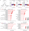
Hypometabolic mismatch with atrophy and tau pathology in mixed Alzheimer's and Lewy body disease
- PMID: 39573823
- PMCID: PMC12073973
- DOI: 10.1093/brain/awae352
Hypometabolic mismatch with atrophy and tau pathology in mixed Alzheimer's and Lewy body disease
Abstract
Polypathology is a major driver of heterogeneity in the clinical presentation and extent of neurodegeneration (N) in patients with Alzheimer's disease (AD). Beyond amyloid (A) and tau (T) pathologies, over half of patients with AD have concomitant pathology such as α-synuclein (S) in mixed AD with Lewy body disease (LBD). Patients with multiple aetiology dementia such as AD + LBD have faster progression and potentially differential responses to targeted treatments, although the diagnosis of AD + LBD can be challenging given the overlapping clinical and imaging features. Development and validation of improved in vivo biomarkers are required to study relationships between N and S and identify imaging patterns reflecting mixed AD + LBD pathologies. We hypothesized that individual proteinopathies, such as T and S, are associated with commensurate levels of N. Thus, we assessed biomarkers of A, T, N and S with PET, MRI and CSF seeding amplification assay (SAA) data to determine molecular presentations of mixed A+S+ versus A+S- cognitively impaired patients from the Alzheimer's Disease Neuroimaging Initiative (ADNI). Strikingly, A+S+ patients had parieto-occipital 18F-fluorodeoxyglucose hypometabolism (a measure of N) disproportionate to the degree of regional atrophy or T burden, highlighting worse hypometabolism associated with S+ status on SAA. Following up on this hypometabolic mismatch with CSF metabolite and proteome analyses, we found that A+S+ patients exhibited lower CSF levels of dopamine metabolites and synaptic markers like neuronal pentraxin-2 (NPTX2), suggesting that altered neurotransmission and neuron integrity contribute to this dissociation between metabolic PET and MRI. Potential confounders exist when studying relations between N, AD and LBD pathologies, including neuroinflammation and other non-Alzheimer's pathologies in mixed dementia, although our findings imply posterior hypometabolic mismatch is related more to S than vascular or TDP-43 pathology. Overall, A+S+ patients had posterior mismatch with excessive 18F-fluorodeoxyglucose hypometabolism relative to atrophy or T load, possibly reflecting impaired neuronal integrity. Further research must disentangle the impact of multiple proteinopathies and clinicopathologic factors on hypometabolism and atrophy. Cumulatively, patients with mixed AD + LBD aetiologies harbour a unique metabolic PET mismatch signature.
Keywords: Alzheimer’s disease; Lewy body; biomarkers; metabolism; multiple aetiology dementia; α-synuclein.
Published by Oxford University Press on behalf of the Guarantors of Brain 2024.
Conflict of interest statement
L.X. received personal consulting fees from Galileo CDS, Inc. L.X. has become an employee of Siemens Healthineers since May 2022 but the current study was conducted during his employment at the University of Pennsylvania. D.A.W. reports grants from Merck, Biogen, Eli Lilly/Avid and additional fees from GE Healthcare, Functional Neuromodulation and Neuronix, all outside this work. I.M.N. reports fees from Biogen outside this work. The remaining authors report no competing interests.
Figures


Similar articles
-
Influence of alpha-synuclein on glucose metabolism in Alzheimer's disease continuum: Analyses of α-synuclein seed amplification assay and FDG-PET.Alzheimers Dement. 2025 Feb;21(2):e14571. doi: 10.1002/alz.14571. Alzheimers Dement. 2025. PMID: 39935389 Free PMC article.
-
Deep learning reveals pathology-confirmed neuroimaging signatures in Alzheimer's, vascular and Lewy body dementias.Brain. 2025 Jun 3;148(6):1963-1977. doi: 10.1093/brain/awae388. Brain. 2025. PMID: 39657969 Free PMC article.
-
Effect of Alzheimer's Disease and Lewy Body Disease on Metabolic Changes.J Alzheimers Dis. 2021;79(4):1471-1487. doi: 10.3233/JAD-201094. J Alzheimers Dis. 2021. PMID: 33459638
-
2014 Update of the Alzheimer's Disease Neuroimaging Initiative: A review of papers published since its inception.Alzheimers Dement. 2015 Jun;11(6):e1-120. doi: 10.1016/j.jalz.2014.11.001. Alzheimers Dement. 2015. PMID: 26073027 Free PMC article. Review.
-
Clinical and diagnostic implications of Alzheimer's disease copathology in Lewy body disease.Brain. 2024 Oct 3;147(10):3325-3343. doi: 10.1093/brain/awae203. Brain. 2024. PMID: 38991041 Review.
Cited by
-
Rock inhibitors in Alzheimer's disease.Front Aging. 2025 Mar 20;6:1547883. doi: 10.3389/fragi.2025.1547883. eCollection 2025. Front Aging. 2025. PMID: 40182055 Free PMC article. Review.
-
Plasma tau biomarkers are distinctly associated with tau tangles and decreased with Lewy body pathology.Alzheimers Dement. 2025 Aug;21(8):e70562. doi: 10.1002/alz.70562. Alzheimers Dement. 2025. PMID: 40791093 Free PMC article.
-
Tau-Clinical Mismatch Identifies Individuals with Co-Pathology and Predicts Clinical Trajectory.medRxiv [Preprint]. 2025 Jul 25:2025.07.25.25332195. doi: 10.1101/2025.07.25.25332195. medRxiv. 2025. PMID: 40778170 Free PMC article. Preprint.
-
Comorbid Pathologies and Their Impact on Dementia with Lewy Bodies-Current View.Int J Mol Sci. 2025 Aug 8;26(16):7674. doi: 10.3390/ijms26167674. Int J Mol Sci. 2025. PMID: 40868991 Free PMC article. Review.
References
-
- Colom-Cadena M, Gelpi E, Charif S, et al. . Confluence of α-synuclein, tau, and β-amyloid pathologies in dementia with Lewy bodies. J Neuropathol Exp Neurol. 2013;72:1203–1212. - PubMed

