. 2025 Jun;12(3):2321-2334.
doi: 10.1002/ehf2.15173. Epub 2024 Nov 21.

Trametinib alters contractility of paediatric Noonan syndrome-associated hypertrophic myocardial tissue slices

Affiliations

Trametinib alters contractility of paediatric Noonan syndrome-associated hypertrophic myocardial tissue slices

Jules Hamers et al. ESC Heart Fail. 2025 Jun.

Abstract

Aims: No curative treatment is available for RASopathy-associated childhood-onset hypertrophic cardiomyopathy (RAS-CM). Preclinical data and individual reports suggest a beneficial effect of small molecules targeting the RAS-mitogen-activated protein (MAP) kinase (MAPK) pathway in severely affected RAS-CM patients. The aim of this study was to evaluate the biophysical effects of trametinib, rapamycin and dasatinib on cultivated myocardial tissue slices of a paediatric RAS-CM patient using biomimetic cultivation chambers (BMCCs) and to correlate the findings with clinical data.

Methods: Contracting right ventricular (RV) tissue slices were prepared from resected myocardium, cultivated in BMCCs and treated with distinct molecules directly and indirectly targeting the RAS-MAPK pathway (trametinib, rapamycin and dasatinib) or dimethyl sulfoxide (DMSO). Tissue biophysical properties were assessed using electrical stimulation protocols. Contractile function, force-frequency relationship and post-pause potentiation were compared before and after treatment. These parameters correlated to L-type Ca2+ channel function and sarcoplasmic Ca2+ loading.

Results: In vivo, off-label treatment with MAPK kinase (MEK) inhibitor trametinib of a child with severe RAS-CM resulted in a modest reduction of RV outflow tract (RVOT) obstruction (RVOT 151 to 122 mmHg after 11 weeks) and improved diastolic function (E/A 0.68 to 1.09 after 11 weeks) and myocardial strain [RV global radial strain (RV-GRS) 25.94 to 42.76; RV global circumferential strain (RV-GCS) -15.26 to -18.61; and RV global longitudinal strain (RV-GLS) -10.31 to -16.78 at 11 weeks], as determined by echocardiography and cardiac magnetic resonance tomography. In cultivated RV myocardial tissue slices, contraction force decreased after addition of trametinib and rapamycin but not after addition of DMSO and dasatinib. Improvement of Ca2+ handling, as depicted by a more positive force-frequency relationship and enhanced post-pause potentiation (31.2%), was noted in the trametinib-treated slice. The increase in post-pause potentiation was less pronounced in rapamycin-treated (26%) and absent in dasatinib-treated (<1%) slices.

Conclusions: Ex vivo analysis of cultivated and electrically stimulated RV myocardial tissue slices of a patient with RAS-CM showed decreased contractility and improved sarcoplasmic reticulum function after addition of trametinib and in part after addition of rapamycin, but not after addition of dasatinib.

Keywords: Noonan; RAF1; cultivation; myocardial slices; trametinib.


Conflict of interest statement

AD is a shareholder of InVitroSys GmbH, which developed the MyoDish cultivation system used for this study. CMW is a consultant for Bristol Myers Squibb, Rocket Pharmaceuticals, Day One Biopharmaceuticals, BioMarin Pharmaceutical, Adrenomed AG and Pliant Therapeutics. CMW has an ownership interest in Preventage Therapeutics.

Figures

Figure 1
Figure 1
Peak dP right ventricular outflow tract (RVOT) and zlog N‐terminal prohormone of brain natriuretic peptide (NT‐proBNP) over time. (A) Decrease of the peak RVOT gradient measured by continuous‐wave Doppler on transthoracic echocardiographies. (B) NT‐proBNP zlog (indicated with ‘•’) and absolute levels (indicated with ‘▪’) at given time points prior to and after the introduction of treatment with the MEK1/2 inhibitor (MEKi) trametinib (indicated with a vertical dotted line).
Figure 2
Figure 2
Cardiac echography before and after treatment with MEK1/2 inhibitor (MEKi) trametinib. (A–F) Echocardiogram prior to treatment with MEKi trametinib. (A, B) Parasternal long‐axis and parasternal short‐axis mid‐ventricle at the papillary muscles showed massive left ventricular hypertrophy (*) with a maximum interventricular septal thickness (IVSd max) of 26 mm (z‐score 7.26) and a maximum left ventricular posterior wall thickness (LVPWd max) of 18 mm (z‐score 6.70). The left ventricular ejection fraction (EF) was hyperdynamic (EF 91%). (C, D) Parasternal short‐axis, base, continuous‐wave Doppler through right ventricular outflow tract (RVOT) depicted a Vmax of 6.14 m/s, corresponding to a peak gradient (dP) of 151 mmHg. (E, F) Four‐chamber view, pulsed‐wave Doppler through mitral valve (MV) (left) and septal tissue Doppler (right) showing diastolic dysfunction with MV E/A 0.68, MV E/e′ 9.5 and e′sept 0.07 m/s. (G–L) Echocardiogram of the same patient 4 months after introduction of treatment with MEKi trametinib. (G, H) Parasternal long‐axis and parasternal short‐axis mid‐ventricle at the papillary muscles showed unchanged massive left ventricular hypertrophy with an IVSd max of 26 mm (z‐score 7.26) and an LVPWd max of 18 mm (z‐score 6.7). The left ventricular EF is hyperdynamic (EF 91%). (I, J) Parasternal short‐axis, base, continuous‐wave Doppler through RVOT depicting Vmax of 5.24 m/s, corresponding to a peak gradient (dP) of 109 mmHg; 5.24 m/s = peak dP RVOT 109 mmHg. (K, L) Four‐chamber view, pulsed‐wave Doppler through MV (left) and septal tissue Doppler (right) showing diastolic dysfunction with MV E/A 1.13, MV E/e′ 10.3 and e′sept 0.06 m/s.
Figure 3
Figure 3
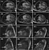
Cardiac magnetic resonance tomography before, 1 week after and 4 months after treatment with MEKi trametinib. (A–D) Cardiac magnetic resonance tomography prior to treatment with MEKi trametinib. (A) Four‐chamber view, end‐diastolic frame. Interventricular septal hypertrophy indicated with ‘*’. (B) Four‐chamber view, end‐systolic frame. Left ventricular hypertrophy indicated with ‘*’. (C) Short‐axis view, end‐diastolic frame. Right ventricular outflow tract obstruction marked with an arrow (➔). (D) Short‐axis view, end‐systolic frame. Right ventricular outflow tract obstruction marked with an arrow (➔). (E–H) Cardiac magnetic resonance tomography after 1 week of treatment with MEKi trametinib. (E) Four‐chamber view, end‐diastolic frame. Interventricular septal hypertrophy indicated with ‘*’. (F) Four‐chamber view, end‐systolic frame. Left ventricular hypertrophy indicated with ‘*’. (G) Short‐axis view, end‐diastolic frame. Right ventricular outflow tract obstruction marked with an arrow (➔). (H) Short‐axis view, end‐systolic frame. Right ventricular outflow tract obstruction marked with an arrow (➔). (I–L) Cardiac magnetic resonance tomography after 11 weeks of treatment with MEKi trametinib. (I) Four‐chamber view, end‐diastolic frame. Interventricular septal hypertrophy indicated with ‘*’. (J) Four‐chamber view, end‐systolic frame. Left ventricular hypertrophy indicated with ‘*’. (K) Short‐axis view, end‐diastolic frame. Right ventricular outflow tract obstruction marked with an arrow (➔). (L) Short‐axis view, end‐systolic frame. Right ventricular outflow tract obstruction marked with an arrow (➔).
Figure 4
Figure 4
Contraction‐force analysis. (A) Average cyclic height of the contraction force. The Y axis visualizes the normalized average contraction force of the slices in %. The contraction was normalized to the contraction strength t = 140. The X axis indicates the cultivation time in hours. The vertical red line indicates the start of the treatments (t = 190), which were continued until the end of cultivation (indicated by the red horizontal arrow). The red triangles (▴) indicate medium changes of the biomimetic cultivation chambers (BMCCs). Because of the influence of the medium change on contractility, the corresponding points have been omitted. All slices were statistically compared to dimethyl sulfoxide (DMSO) 0.1%‐treated control [trametinib (P = 0.023), rapamycin (P < 0.001) and dasatinib (P = 0.066)] (two‐way ANOVA, with Dunnett correction for multiple comparisons, α = 0.05). (B) Medium change did not lead to a contractility depression under trametinib treatment. The Y axis displays the contractility change (%), comparing the absolute contraction force 1 h before and 9 h after medium change. The X axis before and after treatment with the respective substance. Each data point resembles one medium change. Trametinib treatment prevented the contractility depression due to medium change (P = 0.057). Statistical test: Mann–Whitney U test (α = 0.05).
Figure 5
Figure 5
Post‐pause potentiation at a pause duration of 120 s. The Y axis visualizes the difference in potentiation (%) at a pause duration of 120 s of each slice between before and after 8 day treatment. DMSO, dimethyl sulfoxide.
Figure 6
Figure 6
Force–frequency relationship comparison between baseline (i.e., pre‐treatment), 4 days after treatment and 8 days after treatment. Contraction force data points were normalized to the contraction force at 30 b.p.m., as this is the base stimulation frequency of the biomimetic cultivation chambers (BMCCs). Each data point is the average of two assessments performed in a 24 h time frame. Whiskers display the SEM. DMSO, dimethyl sulfoxide.
Figure 7
Figure 7
The RAS–mitogen‐activated protein kinase (MAPK) pathway and the downstream effects in homeostasis and with a RAF1 variant. In homeostasis, growth‐factor receptor (GFR) activation follows the canonical RAS–MAPK pathway, maintaining nominal sarcoplasmic reticulum (SR) function. A gain‐of‐function RAF1 variant, as seen in a portion of the Noonan syndrome associated RASopathy patients, leads to overactivation of RAF1 and Ca2+ leakage from the SR. Based on published literature in combination with our findings, trametinib is thought to mitigate the overactivation of the RAS–MAPK pathway initiated by mutated RAF1. This leads to improved Ca2+ handling by the SR and L‐type calcium channels. RyR, ryanodine receptor; SERCA, sarcoplasmic/endoplasmic reticulum Ca2+‐ATPase.

References

    1. Kaltenecker E, Schleihauf J, Meierhofer C, Shehu N, Mkrtchyan N, Hager A, et al. Long‐term outcomes of childhood onset Noonan compared to sarcomere hypertrophic cardiomyopathy. Cardiovasc Diagn Ther 2019;9:S299‐s309. doi:10.21037/cdt.2019.05.01 - DOI - PMC - PubMed
    1. Roberts AE, Allanson JE, Tartaglia M, Gelb BD. Noonan syndrome. Lancet 2013;381:333‐342. doi:10.1016/S0140-6736(12)61023-X - DOI - PMC - PubMed
    1. Lipshultz SE, Orav EJ, Wilkinson JD, Towbin JA, Messere JE, Lowe AM, et al. Risk stratification at diagnosis for children with hypertrophic cardiomyopathy: an analysis of data from the Pediatric Cardiomyopathy Registry. Lancet 2013;382:1889‐1897. doi:10.1016/S0140-6736(13)61685-2 - DOI - PMC - PubMed
    1. Meier AB, Raj Murthi S, Rawat H, Toepfer CN, Santamaria G, Schmid M, et al. Cell cycle defects underlie childhood‐onset cardiomyopathy associated with Noonan syndrome. iScience 2022;25:103596. doi:10.1016/j.isci.2021.103596 - DOI - PMC - PubMed
    1. Hanses U, Kleinsorge M, Roos L, Yigit G, Li Y, Barbarics B, et al. Intronic CRISPR repair in a preclinical model of Noonan syndrome‐associated cardiomyopathy. Circulation 2020;142:1059‐1076. doi:10.1161/CIRCULATIONAHA.119.044794 - DOI - PubMed

MeSH terms

LinkOut - more resources