

Differential diagnosis of gastrointestinal stromal tumors versus leiomyomas by special stains
- PMID: 39574038
- PMCID: PMC11580362
- DOI: 10.1186/s12876-024-03511-5
Differential diagnosis of gastrointestinal stromal tumors versus leiomyomas by special stains
Abstract
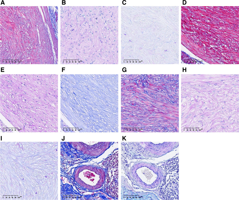
The objective of the study was to investigate whether special stains can differentiate gastrointestinal stromal tumors (GISTs) and gastrointestinal leiomyomas (GILs). In this retrospective study, 39 cases of GISTs (diameter, 0.2-8.8 cm) and 75 cases of GILs (diameter, 0.2-4.5 cm) were recruited, all biopsy specimens were obtained by endoscopic submucosal dissection (ESD) and endoscopic mucosal resection (EMR) excision, and the depth of excision included the whole mucosa, mucosal myometria, and most submucosa. GISTs and GILs were the most common types of mesenchymal tumors found anywhere along the gastrointestinal (GI) tract, from the esophagus to the rectum. GISTs were often associated with a higher risk of malignancy. In this study, the gender, age of onset, size and sites of the lesions, together with the number of mucosal or lamina propria lesions all have significant differences, nevertheless, there was no significant difference in cell morphology of GISTs and GILs tested by hematoxylin eosin (H&E) stain, and all showed low echo areas by EUS examination. In this retrospective study, the GISTs and GILs had been diagnosed by immunohistochemistry combined with clinical morphology. Subsequently, special stains including Masson's trichrome (MT) stain, Alcian blue periodic acid-Schiff (AB-PAS) stain (pH 2.5), Wright-Giemsa (W-G) stain and periodic acid-Schiff (PAS) combined with diastase periodic acid-Schiff (D-PAS) stains were also applied in the diagnosis, the retrospective study results showed that 92.3% GISTs were stained blue with MT stain, 97.3% GILs were stained red with MT stain (P < 0.01), almost all GISTs were PAS-negative (light purple), in contrast, all GILs were PAS-positive (rose red) (P < 0.01), all of these experiments set control using the blood vessels stained by MT and AB-PAS stains. Nevertheless, there was no significant difference between GISTs and GILs stained by W-G stain. These obvious and meaningful differential results were also confirmed in the detection of new GISTs and GILs cases using MT and AB-PAS stains. In conclusion, MT and AB-PAS stains could also identify GISTs and GILs cases, particularly, AB-PAS was more sensitive and more specific, providing a more cost-effective, simple, and high sensitivity and specificity inspection methods, which should be noticed and widely used in the future, especially in resource-limited grass-roots testing institution or in cases with inconclusive immunostains or insufficient material.
Keywords: Gastrointestinal leiomyoma; Gastrointestinal stromal tumor; Malignancy; Special stain.
© 2024. The Author(s).
Conflict of interest statement
Declarations. Ethics approval and consent to participate: All the recruits of this present study were informed and have written informed consent to participate the research, meanwhile, this present study was approved by the ethics committee of Hubei Provincial Hospital of Integrated Chinese and Western Medicine. Consent for publication: All participants were informed and consented to the publication of the results of the study. This present manuscript has not been published or accepted for publication, and the department of pathology, Hubei Provincial Hospital of Integrated Chinese and Western Medicine was fully aware of this submission, and was consent for publication. Competing interests: The authors declare no competing interests.
Figures


References
-
- Munteanu A, Patrascu S, Bordu S, et al. Clinical and Morphological Characteristics of Gastrointestinal Stromal Tumor. Chirurgia (Bucur). 2023;118(6):618–23. - PubMed
MeSH terms
Substances
LinkOut - more resources
Full Text Sources
Miscellaneous
