

Infectious keratoconjunctivitis in European bison (Bison bonasus) in Poland: risk factors, epidemiology and anatomopathological changes with analysis of potential role of Thelazia nematodes in the disease development
- PMID: 39574077
- PMCID: PMC11580673
- DOI: 10.1186/s12917-024-04375-3
Infectious keratoconjunctivitis in European bison (Bison bonasus) in Poland: risk factors, epidemiology and anatomopathological changes with analysis of potential role of Thelazia nematodes in the disease development
Abstract
Background: Infectious keratoconjunctivitis (IKC) is a common ocular disease of ruminants worldwide. Recently, an outbreak of infectious keratoconjunctivitis was observed in the European bison in Poland. Hundreds of animals show conjunctival congestion, corneal opacity, and ulceration, leading to total blindness. The present study aimed to examine the ocular changes of European bison and patterns of the disease occurrence with special emphasis on the role of Thelazia nematodes in the development of IKC.
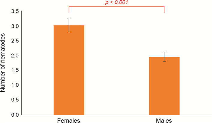
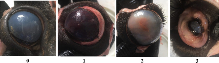
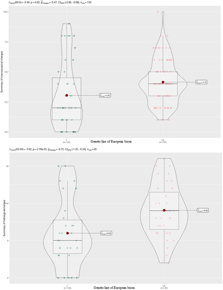
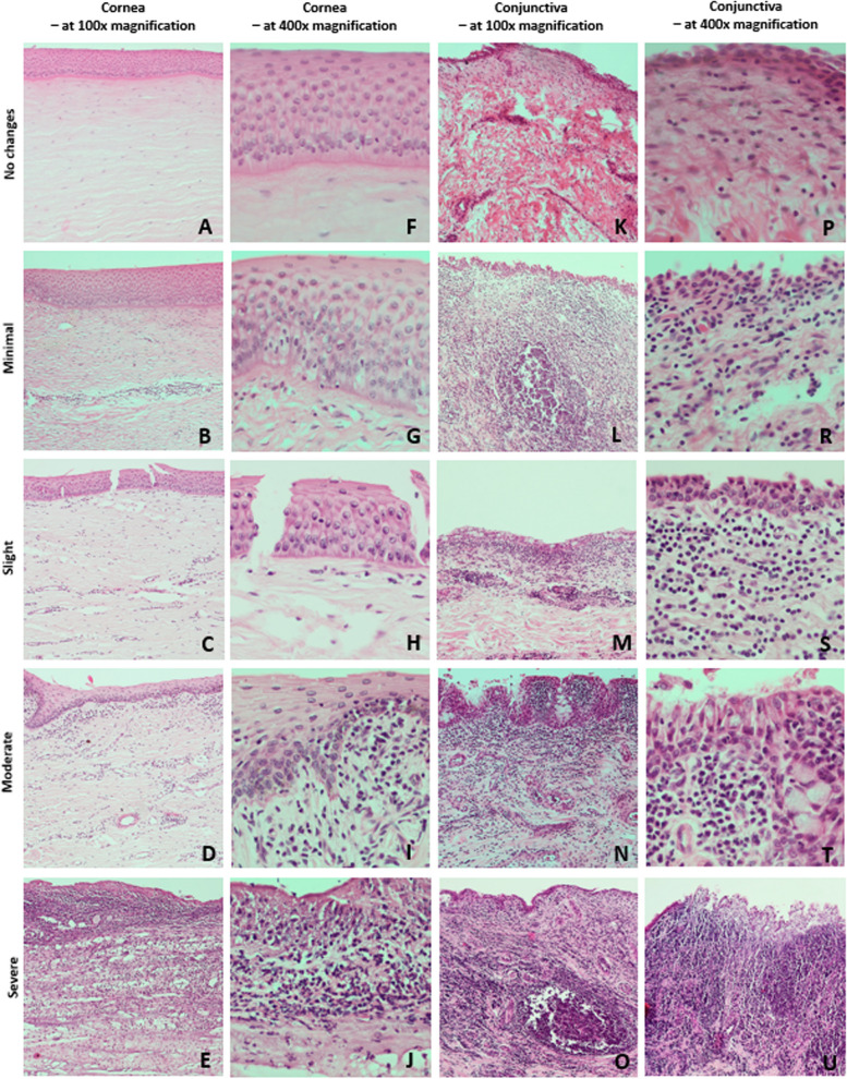
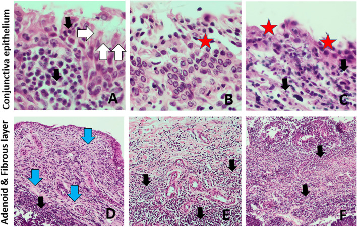
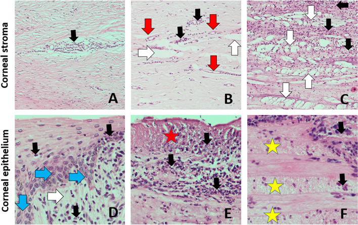
Results: The eyes of 131 European bison, showing ocular signs and clinically healthy, were collected in Poland in 2021 - 2022 and subjected to parasitological and histopathological examination. Histopathology showed varying lesions, including corneal erosions or ulcers, diffuse purulent infiltrates to lymphocytic infiltration in the cornea, and lymphocytic or mixed conjunctivitis with CALT stimulation. The severity of ocular changes was higher in European bison from mountain areas and during the winter season. Two species of Thelazia nematodes - T. skrjabini and T. gulosa have been isolated from eyes. Prevalence of infection reached over 66.4%, and the infection intensity ranged from 1 to 16 nematodes per individual. Although nematodes of the genus Thelazia were prevalent in European bison, their occurrence did not correspond with the severity of ocular changes.
Conclusions: The results of our studies allowed to identify patterns related to the first outbreak of infectious keratoconjunctivitis in European bison. Living in mountain areas and winter season were the most predisposing factors for the development of ocular changes. Despite the high prevalence of Thelazia nematodes in the present study, their role in forming ocular lesions was not confirmed.
Keywords: Bison bonasus; Eyeworm; IKC outbreak; Ocular lesions.
© 2024. The Author(s).
Conflict of interest statement
Declarations. Ethics approval and consent to participate: The authors confirm that the ethical policies of the journal, as noted on the journal’s author guidelines page, have been adhered with accordance to Directive 2010/63/EU of The European Parliament and of The Council of 22 September 2010 on the protection of animals used for scientific purposes. All European bison were legally culled on the basis of permission from the General Directorate for Environmental Protection in Poland. The decision of General Directorate for Environmental Protection was implemented by the Regional Head Offices of National Forests, which supervised animals sacrifice. European bison as wild animals, were culled by professional hunters with respect to the animals welfare and safety rules, in the presence of qualified veterinarians. Consent for publication: Not applicable. Competing interests: The authors declare no competing interests.
Figures
References
-
- Sánchez Romano J, Mørk T, Laaksonen S, Ågren E, Nymo IH, Sunde M, Tryland M. Infectious keratoconjunctivitis in semi-domesticated Eurasian tundra reindeer (Rangifer tarandus tarandus): microbiological study of clinically affected and unaffected animals with special reference to cervid herpesvirus 2. BMC Vet Res. 2018;14:15. - DOI - PMC - PubMed
-
- Degiorgis MP, Frey J, Nicolet J, Abdo EM, Fatzer R, Schlatter Y, et al. An outbreak of infectious keratoconjunctivitis in Alpine chamois (Rupicapra r. rupicapra) in Simmental-Gruyères, Switzerland. SAT Schweiz. Arch Für Tierheilkd. 2000;42:520–7.
MeSH terms
LinkOut - more resources
Full Text Sources
