. 2024 Nov 13;32(12):1881-1890.
doi: 10.32604/or.2024.048007. eCollection 2024.

Caffeic acid hinders the proliferation and migration through inhibition of IL-6 mediated JAK-STAT-3 signaling axis in human prostate cancer

Affiliations

Caffeic acid hinders the proliferation and migration through inhibition of IL-6 mediated JAK-STAT-3 signaling axis in human prostate cancer

Yuan Yin et al. Oncol Res. .

Abstract

Background: Caffeic acid (CA) is considered a promising phytochemical that has inhibited numerous cancer cell proliferation. Therefore, it is gaining increasing attention due to its safe and pharmacological applications. In this study, we investigated the role of CA in inhibiting the Interleukin-6 (IL-6)/Janus kinase (JAK)/Signal transducer and activator of transcription-3 (STAT-3) mediated suppression of the proliferation signaling in human prostate cancer cells.

Materials and methods: The role of CA in proliferation and colony formation abilities was studied using 3-[4,5-dimethylthiazol-2-yl]-2,5 diphenyl tetrazolium bromide (MTT) assay and colony formation assays. Tumour cell death and cell cycle arrest were identified using flow cytometry techniques. CA treatment-associated protein expression of mitogen-activated protein kinase (MAPK) families, IL-6/JAK/STAT-3, proliferation, and apoptosis protein expressions in PC-3 and LNCaP cell lines were measured using Western blot investigation.

Results: We have obtained that treatment with CA inhibits prostate cancer cells (PC-3 and LNCaP) proliferation and induces reactive oxygen species (ROS), cell cycle arrest, and apoptosis cell death in a concentration-dependent manner. Moreover, CA treatment alleviates the expression phosphorylated form of MAPK families, i.e., extracellular signal-regulated kinase 1 (ERK1), c-Jun N-terminal kinase (JNK), and p38 in PC-3 cells. IL-6 mediated JAK/STAT3 expressions regulate the proliferation and antiapoptosis that leads to prostate cancer metastasis and migration. Therefore, to mitigate the expression of IL-6/JAK/STAT-3 is considered an important target for the treatment of prostate cancer. In this study, we have observed that CA inhibits the expression of IL-6, JAK1, and phosphorylated STAT-3 in both PC-3 and LNCaP cells. Due to the inhibitory effect of IL-6/JAK/STAT-3, it resulted in decreased expression of cyclin-D1, cyclin-D2, and CDK1 in both PC-3 cells. In addition, CA induces apoptosis by enhancing the expression of Bax and caspase-3; and decreased expression of Bcl-2 in prostate cancer cells.

Conclusions: Thus, CA might act as a therapeutical application against prostate cancer by targeting the IL-6/JAK/STAT3 signaling axis.

Keywords: Apoptosis; Caffeic acid (CA); Proliferation; Prostate cancer; Signal transducer and activating transcription-3 (STAT-3).


Conflict of interest statement

The authors declare that they have no conflicts of interest to report regarding the present study.

Figures
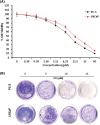
Figure 1
Figure 1. The effect of CA on cell viability and colony formation in prostate cancer cells. (A) Cytotoxicity potential of CA on PC-3 and LNCaP cells. Statistical analysis revealed a significant difference (p < 0.05) in cell viability between the CA-treated group and the control group, indicating the cytotoxic effects of CA on the prostate cancer cells. (B) CA treatment-mediated colony formation was studied by crystal violet staining.
Figure 2
Figure 2. CA enhances reactive oxygen species generation, cell cycle arrest and apoptotic cell death in PC-3 cells. (A) Cells were treated with varying concentrations of CA for a duration of 24 h. Subsequently, they were exposed to DCFH-DA, and ROS was quantified by flow cytometry. (B) CA treatment mediated different phases of the cell cycle was assessed by flow cytometer. Histograms represented the percentages of different cell cycle phases such as G1, S, and G2/M Populations. (C) The role of CA treatment associated with apoptosis of PC-3 cells stained with Annexin V and PI and quantified by flow cytometer. Histograms represented the percentages of apoptosis and viable populations.
Figure 3
Figure 3. CA suppresses phosphorylation of MAPK proteins in prostate cancers. (A) Western blotting images were used to visualize the phosphorylation levels of p38, JNK, and ERK-1 expressions in PC-3 cells. (B) The bar diagram exhibits the mean ± SD of the relative gene expression obtained from three independent experiments. The results show significant differences compared to the control protein β-actin, with a statistical significance level of p < 0.05. Data (a, b, c) are statistically significant when comparing with other groups.
Figure 4
Figure 4. CA attenuates IL-6/JAK/STAT signaling in prostate cancer cell line. (A) Western blotting images were obtained for IL-6, JAK-1, and p-STAT-3 (tyr705) proteins in PC-3 cells. (B) The bar diagram illustrates significant differences in relative protein expression compared to the control protein β-actin. Data has shown to mean ± standard deviation and data (a, b, c) are statistically significant comparing with other groups. (C) Western blotting images were obtained for IL-6, JAK-1, and p-STAT-3 (tyr705) proteins in LNCaP cells. (D) The bar diagram illustrates significant differences in relative protein expression compared to the control protein β-actin. Results indicated by the mean ± SD derived from three independent experimental assays (p < 0.05). Data (a, b, c) are statistically significant when comparing with other groups. (E and F) Western blot data on CA treatment on IL-6 induced expression of, JAK-1, and p-STAT-3 (tyr705) in PC-3 and LNCaP cells.
Figure 5
Figure 5. CA inhibited the proliferation marker expression in PC-3 cells. (A) Western blotting images were obtained for cyclin-D1, CDK-2, and cyclin-D2 protein expressions in PC-3 cells. (B) The bar diagram illustrates significant differences in relative protein expression compared to the control protein β-actin, as indicated by the mean ± SD derived from three independent experimental assays (p < 0.05). Data (a, b, c) are statistically significant when comparing with other groups.
Figure 6
Figure 6. CA treatment enhances apoptosis protein expressions in PC-3 cells. (A) Western blotting images were obtained for Bax, Caspase-3, and Bcl-2 protein expressions in PC-3 cells. (B) The bar diagram illustrates significant differences in relative protein expression compared to the control protein β-actin, as indicated by the mean ± SD derived from three independent experimental assays (p < 0.05). Data (a, b, c) are statistically significant when comparing with other groups.

References

    1. Rawla, P. (2019). Epidemiology of prostate cancer. World Journal of Oncology , 10(2), 63–89. 10.14740/wjon1191; - DOI - PMC - PubMed
    1. Bray, F., Ferlay, J., Soerjomataram, I., Siegel, R. L., Torre, L. A.et al. (2018). Global cancer statistics 2018: GLOBOCAN estimates of incidence and mortality worldwide for 36 cancers in 185 countries. CA: A Cancer Journal for Clinicians , 68(6), 394–424. 10.3322/caac.21492 - DOI - PubMed
    1. Zhu, Y., Wang, H. K., Qu, Y. Y., Ye, D. W. (2015). Prostate cancer in East Asia: Evolving trend over the last decade. Asian Journal of Andrology , 17(1), 48–57. 10.4103/1008-682X.132780; - DOI - PMC - PubMed
    1. Taitt, H. E. (2018). Global trends and prostate cancer: A review of incidence, detection, and mortality as influenced by race, ethnicity, and geographic location. American Journal of Men’s Health , 12(6), 1807–1823. 10.1177/1557988318798279; - DOI - PMC - PubMed
    1. Sekhoacha, M., Riet, K., Motloung, P., Gumenku, L., Adegoke, A.et al. (2022). Prostate cancer review: Genetics, diagnosis, treatment options, and alternative approaches. Molecules , 27(17), 5730. 10.3390/molecules27175730; - DOI - PMC - PubMed

MeSH terms