. 2024 Oct 25;1(3):umae024.
doi: 10.1093/radadv/umae024. eCollection 2024 Sep.

Magnetic particle imaging enables nonradioactive quantitative sentinel lymph node identification: feasibility proof in murine models

Affiliations

Magnetic particle imaging enables nonradioactive quantitative sentinel lymph node identification: feasibility proof in murine models

Olivia C Sehl et al. Radiol Adv. .

Abstract

Background: Sentinel lymph node biopsy (SLNB) is an important cancer diagnostic staging procedure. Conventional SLNB procedures with 99mTc radiotracers and scintigraphy are constrained by tracer half-life and, in some cases, insufficient image resolution. Here, we explore an alternative magnetic (nonradioactive) image-guided SLNB procedure.

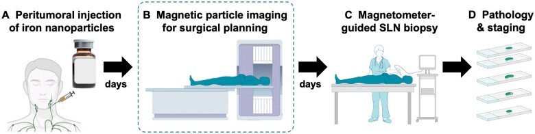
Purpose: To demonstrate that magnetic particle imaging (MPI) lymphography can sensitively, specifically, and quantitatively identify and map sentinel lymph modes (SLNs) in murine models in multiple regional lymphatic basins.

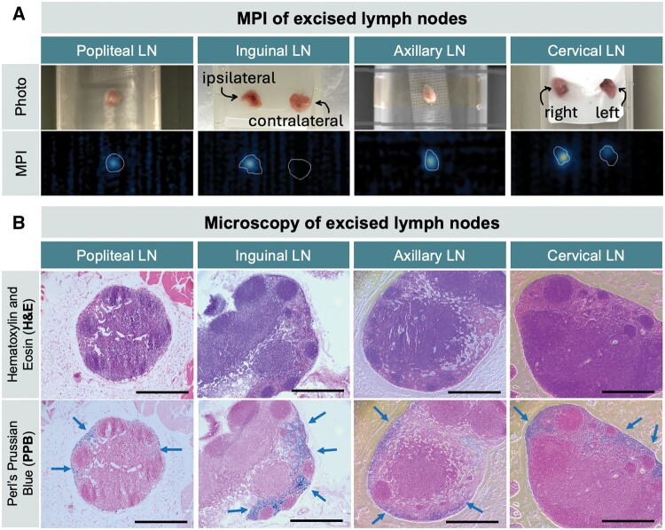
Materials and methods: Iron oxide nanoparticles were administered intradermally to healthy C57BL/6 mice (male, 12-week-old, n = 5). The nanoparticles (0.675 mg Fe/kg) were injected into the tongue, forepaw, base of tail, or hind footpad, then detected by 3-dimensional MPI at multiple timepoints between 1 hour and 4 to 6 days. In this mouse model, the SLN is represented by the first lymph node draining from the injection site. SLNs were extracted to verify the MPI signal ex vivo and processed using Perl's Prussian iron staining. Paired t-test was conducted to compare MPI signal from SLNs in vivo vs. ex vivo and considered significant if P < .05.

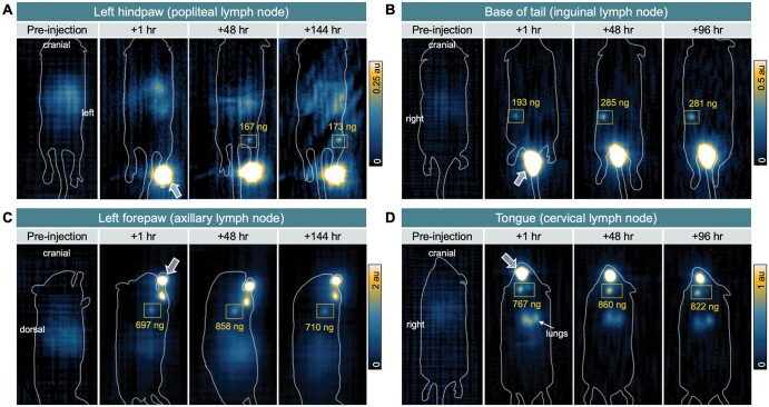
Results: MPI lymphography identified SLNs in multiple lymphatic pathways, including the cervical SLN draining the tongue, axillary SLN draining the forepaw, inguinal SLN draining the tail, and popliteal SLN draining the footpad. MPI signal in lymph nodes was present after 1 hour and stable for the duration of the study (4-6 days). Perl's Prussian iron staining was identified in the subcapsular space of excised SLNs.

Conclusion: Our data support the use of MPI lymphography to specifically detect SLN(s) using a magnetic tracer for a minimum of 4 to 6 days, thereby providing information required to plan the SLN approach in cancer surgery. As clinical-scale MPI is developed, translation will benefit from a history of using iron-oxide nanoparticles in human imaging and recent regulatory-approvals for use in SLNB.

Keywords: iron oxide nanoparticles; lymphography; magnetic particle imaging; pharmacokinetics; quantitative; sentinel lymph node; surgical planning; vivotrax.


Conflict of interest statement

Please see ICMJE form(s) for author conflicts of interest. These have been provided as supplementary materials. Authors O.C. Sehl., K. Guo, A.R. Mohtasebzadeh, P. Kim, B. Fellows, M. Weyhmiller, P.W. Goodwill, and J.M. Greve are employees of Magnetic Insight Inc.

Figures

Figure 1.
Figure 1.
Magnetic particle imaging (MPI) shows in vivo pharmacokinetics of iron oxide nanoparticles to sentinel lymph nodes (SLN) of C57 immunocompetent mice. MPI was acquired preinjection to assess mouse background signal then at 3 timepoints following intradermal injection of VivoTrax to the (A) left hindpaw, (B) right base of tail, (C) left forepaw, and (D) tongue. Arrows indicate location of injection sites. Sentinel lymph nodes are outlined by squares, including the (A) ipsilateral popliteal lymph node, (B) inguinal lymph node, (C) axillary lymph node, and (D) cervical lymph node(s), respectively. Absolute quantification of iron at each lymph node is indicated. A, B, and D are coronal views, whereas imaging of the forepaw required positioning the mouse on its side, resulting in a sagittal view.
Figure 2.
Figure 2.
Measurements of distance and pharmacokinetics of iron from injection site to draining lymph nodes, corresponding with magnetic particle imaging (MPI) data in Figure 1. (A) Cartoon showing lymphatic basins draining from the mouse tongue, forepaw, base of tail, and footpad, to the cervical, axillary, inguinal, and popliteal lymph nodes, respectively. The average distance between the injection site and draining lymph nodes, measured from MPI images, is reported (mean ± standard deviation). (B) Longitudinal quantification of MPI signal at the injection site and SLNs for each lymphatic pathway, expressed as percent injected dose (% ID), from 1 hour up to 96 or 144 hours.
Figure 3.
Figure 3.
Validation for excised lymph nodes by MPI and histology. (A) MPI of excised tissue verifies that in vivo MPI signal originates from draining lymph nodes. (B) Ex vivo histology of excised popliteal, inguinal, axillary, and cervical lymph nodes including adjacent histopathologic slices with hematoxylin-eosin (H&E) staining (top) and Perl’s Prussian blue (PPB) staining (bottom). Iron staining is identified at the periphery of lymph nodes in the subcapsular spaces. Magnification = 10×, scale bars = 0.5 mm.
Figure 4.
Figure 4.
Assessment of preclinical magnetic particle imaging (MPI) technical specifications for sentinel lymph node imaging. (A) Sensitivity and linearity MPI phantom. Three-dimensional MPI of 10 vials with Fe quantities of 25, 50, 75, 100, 150, 200, 300, 400, 600, 800 ng in 7 µL, displayed with window/level = 1/0.5 au. The white dotted line represents the position of the 25-ng VivoTrax sample, which did not meet MPI detection criteria. The limit of detection was determined as 50 ng. Line profiles drawn through MPI signal peaks show signal intensities of each sample and integrated MPI signal is directly linear with Fe quantity (R2 = 0.99). (B) Shine-through/resolution MPI phantom. Three-dimensional MPI of a center vial containing 13.5 µg Fe and satellite vials of 675 ng are 2.5 mm to 20 mm from the source (well center-to-center distance). Phantom wells were 1.5 mm in diameter. MPI is displayed with window/level = 8/4 au. Line plots through MPI signals show that a 675-ng sample can be resolved at 3.5 mm edge-to-edge distance from center vial (5 mm center-to-center distance). Red asterisk (*) indicates the limit of detection (A) and the limit of resolution (B).
Figure 5.
Figure 5.
Proposed clinical all-magnetic sentinel lymph node biopsy (SLNB) workflow. (A) First, an iron oxide nanoparticle tracer is administered adjacent to a tumor. (B) A magnetic particle imaging (MPI) scan is acquired before surgery to identify the location and number of SLNs draining from a primary tumor. Provided the data from this study, this image could be acquired hours to days after iron injection, which provides tremendous clinical flexibility. (C) As currently practiced, the surgery could be guided by a magnetometer in real time and (D) excised SLNs would be sent for pathology and staging.

References

    1. de Bree R, de Keizer B, Civantos FJ, et al.What is the role of sentinel lymph node biopsy in the management of oral cancer in 2020? Eur Arch Otorhinolaryngol. 2021;278(9):3181-3191. - PMC - PubMed
    1. Mahieu R, de Maar JS, Nieuwenhuis ER, et al.New developments in imaging for sentinel lymph node biopsy in early-stage oral cavity squamous cell carcinoma. Cancers (Basel). 2020;12(10):3055. - PMC - PubMed
    1. Parrett BM, Kashani-Sabet M, Singer MI, et al.Long-term prognosis and significance of the sentinel lymph node in head and neck melanoma. Otolaryngol Head Neck Surg. 2012;147(4):699-706. - PubMed
    1. Bluemel C, Herrmann K, Giammarile F, et al.EANM practice guidelines for lymphoscintigraphy and sentinel lymph node biopsy in melanoma. Eur J Nucl Med Mol Imaging. 2015;42(11):1750-1766. - PubMed
    1. de Veij Mestdagh PD, Schreuder WH, Vogel WV, et al.Mapping of sentinel lymph node drainage using SPECT/CT to tailor elective nodal irradiation in head and neck cancer patients (SUSPECT-2): a single-center prospective trial. BMC Cancer. 2019;19(1):1110-1119. - PMC - PubMed

LinkOut - more resources