[Preprint]. 2024 Nov 4:2024.11.04.621790.
doi: 10.1101/2024.11.04.621790.

Constitutive activity of an atypical chemokine receptor revealed by inverse agonistic nanobodies

Affiliations

Constitutive activity of an atypical chemokine receptor revealed by inverse agonistic nanobodies

Claudia V Perez Almeria et al. bioRxiv. .

Abstract

Chemokine stimulation of atypical chemokine receptor 3 (ACKR3) does not activate G proteins but recruits arrestins. It is a chemokine scavenger that indirectly influences responses by restricting the availability of CXCL12, an agonist shared with the canonical receptor CXCR4. ACKR3 is upregulated in numerous disorders. Due to limited insights in chemokine-activated ACKR3 signaling, it is unclear how ACKR3 contributes to pathological phenotypes. One explanation may be that high constitutive activity of ACKR3 drives non-canonical signaling through a basal receptor state. Here we characterize the constitutive action of ACKR3 using novel inverse agonistic nanobodies to suppress basal activity. These new tools promote an inactive receptor conformation which decreased arrestin engagement and inhibited constitutive internalization. Basal, non-chemotactic, breast cancer cell motility was also suppressed, suggesting a role for ACKR3 in this process. The basal receptor activity in pathophysiology may provide a new therapeutic approach for targeting ACKR3.


Conflict of interest statement

Competing interests B.F.V. has an ownership interest in Protein Foundry, L.L.C. and XLock Biosciences, Inc. R.H. is affiliated with QVQ Holding BV. All other authors declare no competing interests.

Figures
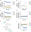
Figure 1.
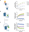
Figure 1.. ACKR3 nanobodies suppress basal β-arrestin2 recruitment.
A-B) Recruitment of β-arr2-mV to ACKR3-Nluc measured by BRET (A) Time-dependent change in BRET over 60 min with either 316 nM of CXCL12 (blue circle) or 1 μM of nanobody (VUN700 (green triangle), VUN701 (purple inverted triangle), VUN702 (yellow diamond)) and (B) dose response curves of CXCL12 or nanobodies at 60 min recorded at 37°C in HEK293T cells. C-D) β-arrestin2 recruitment measured by BRET in agonist mode between donor ACKR3-Nluc and β-arr2-mV in (C) parental HEK293 or in (D) GRK2/3/5/6 KO HEK293 cells. E) Schematic illustration of BRET-based FlAsH-tagged (CCPGCC) sensor F5 (between residues 156 and 157) on β-arrestin2. F) Time-resolved changes in the NanoBRET β-arrestin2 conformational biosensor F5 signal upon ACKR3 activation, following the addition of 316nM of CXCL12 or 1 μM of VUN700, VUN701 or VUN702 at 37°C in parental HEK293 cells. Data are shown as the average ± SD of three independent experiments performed in technical triplicates.
Figure 2.
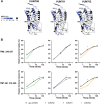
Figure 2.. NMR-based structural characterization of ACKR3 upon nanobodies VUN700 binding reveals a relatively more pronounced “OFF” state of ACKR3 than VUN701-bound state.
A) ACKR3 structure (7SK6 PDB) with NMR peaks M1383×46 and M2125×39 depicted. B) Overlay of M2125×39 peaks from all nanobodies and CXCL12 ligand-bound ACKR3 complexes. C) Overlay of M1383×46 peaks in all nanobodies and CXCL12 ligand-bound states. Upfield peak positions (1H: ~1.3 ppm) of M1383×46 among agonist-bound states supports ring-current shifts due to aromatic side chain interactions.
Figure 3.
Figure 3.. Conformational changes in ACKR3 induced by nanobody binding.
A). Structural representation of the % differential relative fractional uptake (ΔRFU) data (apo ACKR3 – Nb-bound ACKR3) mapped onto the cryo-EM structure of ACKR3 (PDB:7SK5). This depicts reproducible and statistically significant ΔHDX over 120 minutes deuteration. The degree of ΔHDX (% ΔRFU) ΔRFU is represented according to the color scale. Black regions represent those with no sequence coverage. B) Deuterium uptake plots showing time-dependent change in RFU for ACKR3 peptides on the intracellular side upon nanobodies binding, compared to apo ACKR3 (in black). Uptake represents the average and SD of three technical replicates from one biological preparation of ACKR3. Data is representative of three biological replicates. Statistically significant changes were determined using Deuteros 2.0 software (*, p ≤ 0.01).
Figure 4.
Figure 4.. ACKR3 nanobodies differentially change the localization of ACKR3 by capturing ACKR3 at the membrane.
A) Surface ACKR3 detected by flow cytometry upon 316 nM of VUN700 (green triangle), VUN701 (purple inverted triangle) or VUN702 (yellow diamond), or 100 nM CXCL12 (blue circle) over 60 min at 37°C in HEK293 cells. B) Time-dependent internalization measured by BRET, between donor ACKR3-Nluc with mV-CAAX over 60 min with either 316nM of CXCL12 or 1μM of VUN700, VUN701 or VUN702 at 37°C in HEK293T cells. C) Time-dependent ACKR3 localized in the early endosomes measured by BRET, between donor ACKR3-Nluc with Rab5a-mV, over 60 min with either 316nM of CXCL12 or 1μM of VUN700, VUN701 or VUN702 at 37°C in HEK293T cells. Data is shown as the average ± SD of at least three independent experiments performed in duplicates or triplicates. One-way ANOVA, multiple comparisons Dunnett test (* p<0.05).
Figure 5.
Figure 5.. ACKR3 nanobodies mediate GRK-independent and dependent constitutive internalization.
A-D) Internalization of ACKR3-Nluc measured by BRET to the PM probe mV-CAAX in (A-B) parental HEK293 or in (C-D) GRK2/3/5/6 KO HEK293 cells at 37°C. (A, C) Time-dependent change in BRET over 60 min with either 316 nM of CXCL12 (blue circle) or 1 μM of VUN700 (green triangle), VUN701 (purple inverted triangle) or VUN702 (yellow diamond) and (B, D) dose response curves of CXCL12 or nanobodies at 60 min. Data is shown as the average ± SD of four independent experiments performed in triplicate.
Figure 6.
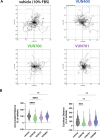
Figure 6.. Basal motility in metastatic breast cancer cells is reduced upon inverse agonist nanobody targeting ACKR3.
A) Representative basal motility paths of MDA-MB-231 metastatic breast cancer cells for 16h in 10% FBS, vehicle condition (in black) or with 100nM of VUN700 (in green), VUN701 (in purple) or VUN400 (in blue). B) Accumulated distance (total distance traveled) and Euclidean distance (Start to end point distance) for individual cells from each of the conditions in A. The positions of at least 120 cells, selected across three replicates (~40 cells/repeat), were tracked and normalized to the vehicle condition. Significance was determined by one-way ANOVA Dunnett test (p<0.0005 (***), <0.0001 (****)).
Figure 7.
Figure 7.. Constitutive activity of atypical chemokine receptor revealed by inverse agonistic nanobodies.
Apo-ACKR3 is constitutively active, which leads to the receptor basal GRK phosphorylation, arrestin recruitment, and internalization. Stimulation with an agonist like CXCL12 fully activates ACKR3 and drives robust phosphorylation, arrestin complexing, and endocytosis. Inverse agonistic nanobodies suppress the constitutive activity of ACKR3, trapping the receptors at the cell surface and reducing interactions with arrestins or GRKs, thereby allowing GRKs and arrestins to be available for other GPCRs. These nanobodies also slow basal cell migration, suggesting a role for ACKR3 constitutive activity in cell motility.

References

    1. Rajagopal S. et al. β-Arrestin- But not G protein-mediated signaling by the “decoy” receptor CXCR7. Proceedings of the National Academy of Sciences of the United States of America 107, 628–632 (2010). 10.1073/pnas.0912852107 - DOI - PMC - PubMed
    1. Ödemis V. et al. The presumed atypical chemokine receptor CXCR7 signals through Gi/o proteins in primary rodent astrocytes and human glioma cells. Glia 60, 372–381 (2012). 10.1002/glia.22271 - DOI - PubMed
    1. Fumagalli A. et al. The atypical chemokine receptor 3 interacts with Connexin 43 inhibiting astrocytic gap junctional intercellular communication. Nature Communications 10.1038/s41467-020-18634-y - DOI - PMC - PubMed
    1. Saaber F. et al. ACKR3 Regulation of Neuronal Migration Requires ACKR3 Phosphorylation, but Not beta-Arrestin. Cell Rep 26, 1473–1488 e1479 (2019). 10.1016/j.celrep.2019.01.049 - DOI - PubMed
    1. Schafer C. T., Chen Q., Tesmer J. J. G. & Handel T. M. Atypical Chemokine Receptor 3 ‘Senses’ CXC Chemokine Receptor 4 Activation Through GPCR Kinase Phosphorylation. Mol Pharmacol (2023). 10.1124/molpharm.123.000710 - DOI - PMC - PubMed

Publication types

LinkOut - more resources