[Preprint]. 2024 Nov 8:2024.11.06.622387.
doi: 10.1101/2024.11.06.622387.

An mRNA Display Approach for Covalent Targeting of a Staphylococcus aureus Virulence Factor

Affiliations

An mRNA Display Approach for Covalent Targeting of a Staphylococcus aureus Virulence Factor

Sijie Wang et al. bioRxiv. .

Update in

Abstract

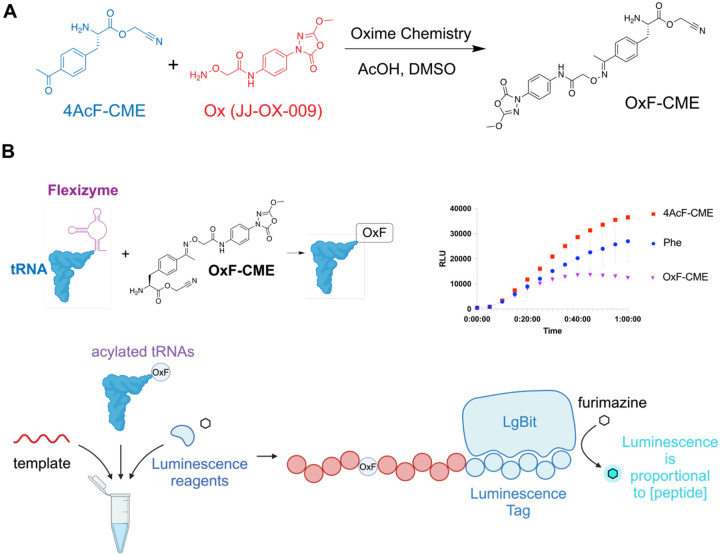
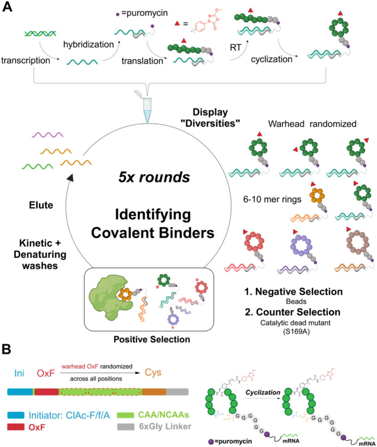
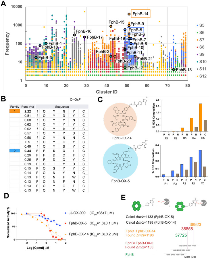
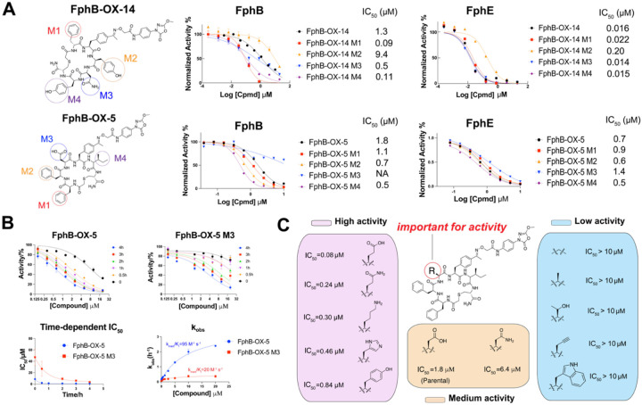
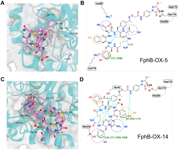
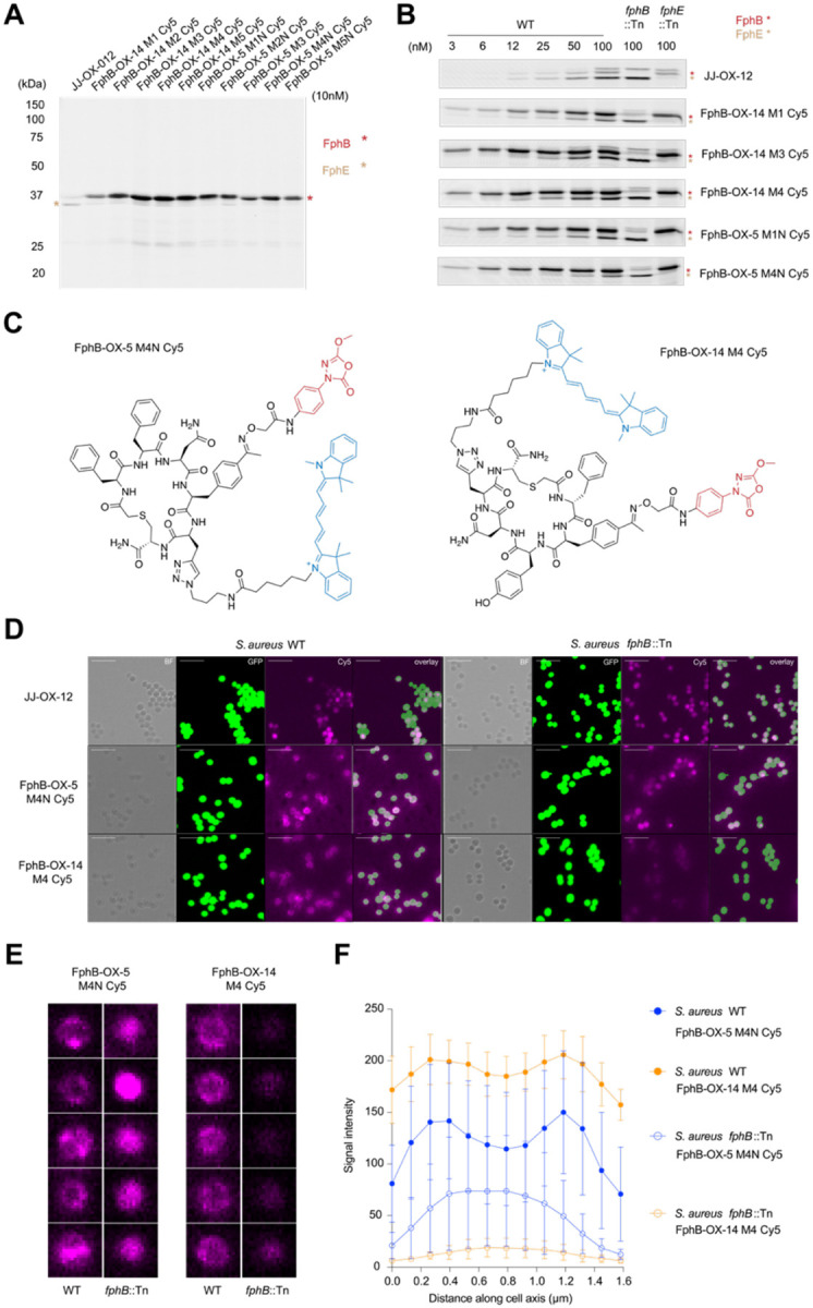
Staphylococcus aureus (S. aureus) is an opportunistic human pathogen that causes over one million deaths around the world each year. We recently identified a family of serine hydrolases termed fluorophosphonate binding hydrolases (Fphs) that play important roles in lipid metabolism and colonization of a host. Because many of these enzymes are only expressed in Staphylococcus bacteria, they are valuable targets for diagnostics and therapeutics. Here we developed and screened highly diverse cyclic peptide libraries using mRNA display with a genetically encoded oxadiazolone (Ox) electrophile that was previously shown to potently and covalently inhibit multiple Fph enzymes. By performing multiple rounds of counter selections with WT and catalytic dead FphB, we were able to tune the selectivity of the resulting selected cyclic peptides containing the Ox residue towards the desired target. From our mRNA display hits, we developed potent and selective fluorescent probes that label the active site of FphB at single digit nanomolar concentrations in live S. aureus bacteria. Taken together, this work demonstrates the potential of using direct genetically encoded electrophiles for mRNA display of covalent binding ligands and identifies potent new probes for FphB that have the potential to be used for diagnostic and therapeutic applications.


Figures

Figure 1.
Figure 1.
Design strategy of covalent mRNA display libraries by genetic encoding of the oxadiazolone electrophile. (A) Synthesis of 4-acetyl-phenylalanine cyanomethyl ester (OxF-CME). The parent oxadiazolone electrophile fragment containing a hydroxylamine was conjugated to the cyanomethyl ester of 4-acyl-phenylalanine (4AcF-CME) using oxime chemistry to obtain the final oxadiazolone phenylalanine cyanomethyl ester (OxF-CME) used for flexizyme-mediated acylation of tRNA. The synthesis of this compound is described in the Supporting Information. (B) Schematic of the NanoBiT luminescence-based assay to evaluate the in vitro translation (IVT) efficiency of non-natural amino acid oxadiazolone phenylalanine (OxF). The modified OxF-CME was used to acylate a tRNA using the flexizyme system followed by in vitro translation of a peptide containing the OxF amino acid and a luminescent peptide tag. Addition of luminescence reagents allows real-time monitoring of peptide translation. Plot shows the resulting translation rates for the natural Phe (phenylalanine; blue), compared to 4Acetyl phenylalanine (4AcF-CME; red), and the oxadiazolone modified phenylalanine (OxF-CME; purple).
Figure 2.
Figure 2.
Synthesis and selections of highly diverse mRNA display libraries with a genetically encoded oxadiazolone warhead to FphB (A) The mRNA libraries were prepared following five main steps (1) DNA transcription to mRNA (2) mRNA templates hybridized to a puromycin linked cDNA (3) flexible in vitro translation and cyclization of peptides from mRNA template (4) reverse transcription of mRNA to generate a cDNA strand. After library synthesis, highly diverse libraries (6-10mer ring sizes with warhead randomized at all positions) were selected against WT rFphB as positive selection, and catalytic dead mutant rFphB (S169A) and beads as negative selections. Different washing stringencies (kinetic long-time washes and 5M guanidine washes) were applied at various rounds of selections. DNA elutes after each round of selection were reamplified and prepared for the next round of selection. (B) Each peptide in the library is composed of N terminal initiator ClAc-R (R=F/f/A; blue) encoded by ATG, variable region (green) and C-terminal cysteine (orange) flanked with a six-glycine linker (grey). In the variable regions, a single warhead OxF encode by CAG was used at all positions. Other canonical amino acids (CAA) and non-canonical amino acids (NCAA) were fully randomized at all other positions using the NNT codon. Peptides were automatically cyclized post translationally through the reaction of the N-terminal chloroacetylated amino acid with the single cysteine side chain thiol in each peptide.
Figure 3.
Figure 3.
Family cluster profiling and hits from mRNA display screening to FphB. (A) Cluster analysis based on atomic similarity of enriched peptide hits. The top 21 hits selected for validation studies are circled in black in the cluster, with FphB-OX-5 and FphB-OX-14 highlighted. S5-12 represents different libraries with different codon tables (see Figure S3 for codon use). S5: 6-8mers, codon table 1; S6: 9-10mers, codon table 1; S7: 6-8mers, codon table 2; S8: 9-10mers, codon table 2; S9: 6-8mers, codon table 3; S10: 9-10mers, codon table 3; S11: 6-8mers, codon table 4; S12: 9-10mers, codon table 4. (B) Table showing sequences and enrichment of two clusters (O=OxF warhead, lowercase letters are d-amino acids, first reside is N-chloroacetyl amino acid). The percentage (Perc. (%)) of DNA copy numbers of each unique macrocycle in total reads of the NGS pool is shown. (C) Structures of the top two lead molecules and their enrichment profile in positive and negative selections in all five rounds. Round 1 uses only positive selection, rounds 2-4, beads were used as negative selections (annotated as N), round 5, S169A catalytic dead mutant rFphB was used for counter selection (annotated as C). (D) IC50 quantifications of JJ-OX-009, FphB-OX-5 and FphB-OX-14 for rFphB (100 nM), measured by the cleavage of the 4-MUB substrate (20 μM). The activity was normalized to a DMSO control, with points and bars representing means ± standard deviation (n=4). (E) Intact rFphB labeling with FphB-OX-5 and FphB-OX-14, validated by deconvoluted mass spectra of rFphB before (green) and after (OX-5: red, OX-14: orange) treatment. MS deconvolution analysis confirmed a single covalent modification of the protein.
Figure 4:
Figure 4:
Structure-dependent activity validations of hits. (A) Each amino acid (highlighted as M1/M2/M3/M4) of FphB-OX-14 and FphB-OX-5 was individually replaced with propargylglycine to generate positional scanning alkyne mutants. Inhibitory activity of alkyne mutants with parental molecules to rFphB were quantified. (B) Time dependent enzymatic activities of OX-5 and OX-5 M3 to rFphB were quantified at six time points (0, 0.5, 1, 2, 3, and 4 hours). Second order rate constants kinact/Ki were determined using time-dependent IC50 values. (C) Structure-activity relationship (SAR) data for FphB-OX-5 at the M3 position. The indicated amino acids were used to individually replace the aspartic acid in the parental structure. The IC50 values of each compound in enzymatic assays against rFphB are listed for each compound.
Figure 5.
Figure 5.
Covalent docking of FphB-OX-5 and FphB-OX-14 to FphB. (A) FphB-OX-5 simulated binding pose modeled in predicted FphB structure. Hydrogen bonds and reactive interaction with S176 shown as gold pseudobonds. (B) Interaction diagram for FphB-OX-5. Hydrogen bonds shown as cyan dashed lines. Van der Waals interactions shown by green arc. (C) FphB-OX-14 simulated binding pose modeled in predicted FphB structure. Hydrogen bonds and reactive interaction with S176 shown as gold pseudobonds. (D) Interaction diagram for FphB-OX-14. Hydrogen bonds shown as cyan dashed lines. Van der Waals interactions shown by green arc. T-stacking interaction shown as black dashed line.
Figure 6.
Figure 6.
Live S. aureus labeling and microscope confocal imaging with fluorescent imaging probes. (A) SDS-PAGE image of live USA300 S. aureus cells (both wild-type and fphB transposon (fphB::Tn) mutant strains)) labeled with 10 nM of JJ-OX-012, as well as the FphB-OX-14 and FphB-OX-5 alkyne scanning mutants. Cells were treated with probes at 37 °C for 1 h before cell lysis, click chemistry labeling with Cy5-azide, SDS-PAGE and fluorescence imaging. (B) Dose-dependent labeling of USA300 S. aureus cells with the indicated probes at concentrations from 3 nM to 100 nM. The location of the FphB protein is shown in red, FphE shown in beige. The fphB transposon mutant strain (fphB::Tn) and fphE transposon mutant strain (fphE::Tn) are shown at the 100 nM probe concentration. (C) Structures of the two fluorescent probes FphB-OX-14 M4 Cy5 and FphB-OX-5 M4(N) Cy5. (D) Confocal micrographs of stationary phase S.aureus USA300-GFP cells labeled with 100 nM JJ-OX-12, FphB-OX-5 M4(N) Cy5 and FphB-OX-14 M4 Cy5. Panel 1:bright-field (BF); panel 2:GFP imaging; panel 3:Cy5 imaging; panel 4:overlay of panels GFP and Cy5. Scale bar: 10 μm (E) Representative single-cell confocal images of WT S. aureus USA300 (left) and fphB transposon (fphB::Tn) mutant cells (right) treated with FphB-OX-5-M4(N) Cy5 and FphB-OX-14 M4 Cy5. Scale bar: 1 μm. (F) Quantified average fluorescence intensity distribution across the cell for each probe in WT and transposon mutant cells. Microscopy images of cells were analyzed by implementing an agile script. The image was converted to a binary format, cells were identified, and the major axis length along with the mean intensity of each cell was calculated. The mean intensity was plotted against the major axis length for each identified cell in the images. Bars represent means ± standard deviation (n =6).

Similar articles

References

    1. Singh J.; Petter R. C.; Baillie T. A.; Whitty A. The Resurgence of Covalent Drugs. Nat Rev Drug Discov 2011, 10 (4), 307–317. 10.1038/nrd3410. - DOI - PubMed
    1. Boike L.; Henning N. J.; Nomura D. K. Advances in Covalent Drug Discovery. Nat Rev Drug Discov 2022, 21 (12), 881–898. 10.1038/s41573-022-00542-z. - DOI - PMC - PubMed
    1. Baillie T. A. Targeted Covalent Inhibitors for Drug Design. Angewandte Chemie International Edition 2016, 55 (43), 13408–13421. 10.1002/anie.201601091. - DOI - PubMed
    1. Ghosh A. K.; Samanta I.; Mondal A.; Liu W. R. Covalent Inhibition in Drug Discovery. ChemMedChem 2019, 14 (9), 889–906. 10.1002/cmdc.201900107. - DOI - PMC - PubMed
    1. Schwartz P. A.; Kuzmic P.; Solowiej J.; Bergqvist S.; Bolanos B.; Almaden C.; Nagata A.; Ryan K.; Feng J.; Dalvie D.; Kath J. C.; Xu M.; Wani R.; Murray B. W. Covalent EGFR Inhibitor Analysis Reveals Importance of Reversible Interactions to Potency and Mechanisms of Drug Resistance. Proceedings of the National Academy of Sciences 2014, 111 (1), 173–178. 10.1073/pnas.1313733111. - DOI - PMC - PubMed

Publication types

LinkOut - more resources