[Preprint]. 2024 Nov 4:2024.11.04.621904.
doi: 10.1101/2024.11.04.621904.

Antigen-Specific T Cell Receptor Discovery for Treating Progressive Multifocal Leukoencephalopathy

Affiliations

Antigen-Specific T Cell Receptor Discovery for Treating Progressive Multifocal Leukoencephalopathy

Sasha Gupta et al. bioRxiv. .

Abstract

Background: Progressive multifocal leukoencephalopathy (PML) is a frequently fatal disease of the central nervous system caused by JC virus (JCV). Survival is dependent on early diagnosis and ability to re-establish anti-viral T cell immunity. Adoptive transfer of polyomavirus-specific T cells has shown promise; however, there are no readily available HLA-matched anti-viral T cells to facilitate rapid treatment.

Objective: Identify epitopes of the JCV major capsid protein VP1 that elicit an immune response in the context of human leukocyte antigen allele A*02:01 (HLA-A2) and isolate cognate T cell receptors (TCRs) from healthy donors. Evaluate individual VP1-specific TCRs for their capacity to be expressed in T cells and clear JCV in vitro.

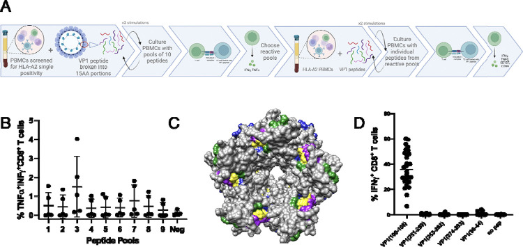
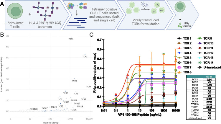
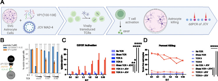
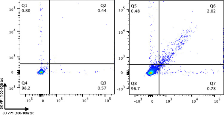
Methods: PBMCs from HLA-A2+ healthy donors were stimulated with peptide libraries tiled across the JCV VP1 protein. Multiple rounds of stimulation were performed to identify the antigens that induced the largest expansion and CD8+ T cell response (measured as INFγ, TNFα, CD137, and CD69 expression). High-affinity, antigen-specific CD8+ T cells were isolated based on intensity of tetramer binding for downstream single-cell TCR sequencing. Candidate TCRs were selected based on tetramer binding affinity and activation assays. Promising TCRs were introduced into the T cell genome via viral transduction for in vitro validation including peptide-pulsed K562 cells and astrocyte cells, and JCV-infected astrocytes.

Results: Four conserved JCV VP1 epitopes (amino acids 100-108, 251-259, 253-262, and 274-283) presented by HLA-A2 were identified. VP1(100-108) consistently elicited the highest level of IFN-γ production from multiple donors and this peptide is in a highly conserved region of VP1. We next identified fourteen high avidity TCRs specific for VP1(100-108). When virally transduced into primary human T cells, seven of these TCRs demonstrated specific binding to VP1(100-108):HLA-A2 tetramers, and four showed increased IFN-γ response when incubated with peptide. Primary CD8+ T cells expressing two of these TCRs cleared both HLA-A2 positive K562 cells and HLA-A2 positive SVG astrocyte cell line presenting exogenously added VP1 peptide at a range of E:T ratios. In addition, both TCR-transduced T cell populations effectively lysed JCV-infected astrocytes.

Conclusions: We identified JCV VP1 epitopes that are immunogenic in the context of HLA-A2 MHC-I, including epitopes that have not been previously described. The VP1(100-108) epitope was used to isolate HLA-A2-restricted TCRs. When cloned into primary human CD8+ T cells, these TCRs recognized VP1 (100-108)-presenting targets, and the transduced T cells conferred cytotoxic activity and eliminated K562 and astrocyte cells displaying the VP1(100-108) peptide and not sham peptide, as well as JCV-infected astrocytes. Taken together, these data suggest that JCV VP1-specific TCRs could be appealing therapeutics for HLA-A2+ individuals with PML in whom intrinsic T cell immunity cannot be rescued.


Figures

Figure 1:
Figure 1:. Defining immunogenic peptide.
A. Schematic of experimental layout. B. CD8+ T cells from 5 donors stimulated 3x with pools of 10 peptides, except pool 9 which had 6 peptides. C. 3D modeling of VP1 pentamer highlighting epitopes (Green: VP1 (100–108), Yellow: VP1 (251–259), (253–262), (274–283), Purple: Published epitopes, Blue: Predicted epitopes from IEDB. D. CD8+ T cells from 3 donors, 10 cell lines each, stimulated 3x with pools of peptides and then individual T2 IFN-γ response.
Figure 2:
Figure 2:. JCV VP1 (100–108) specific T cell receptors (TCR).
A. Schematic of experimental layout. B. Pooled CD8+ T cell lines from 3 healthy donors were split into three different fractions and stained with low, intermediate, or high tetramer concentration. From samples stained with low and intermediate tetramer concentration, live CD3+ CD8+ T cells were sorted from the 90th percentile tetramer(+) gates. Shown here is the cumulative clone count in the 90th percentile gates (y-axis) plotted against maximum enrichment score out of the 11 calculated for each clone (x-axis). Symbol size corresponds to clone count in tetramer-negative gate. Symbol color indicates clone abundance in the single cell TCR dataset (low tetramer concentration condition). High affinity TCRs have high cumulative clone count, high maximum enrichment score, and small symbol size. SumSort = total number in top 10% tetramer binder gate (i.e. 90th percentile) in the 1:2000 + 1:4000 tetramer staining condition. MaxFold = greatest value for fold enrichment (out of the calculated 11). C. Top 14 T cell receptors were virally transduced into human primary T cells in 2 donors. TCR-transduced cells were stimulated with different amounts of VP1(100–108) peptide and tested for intracellular INF-γ content as ratio of positive control (max). EC50 (M) scores populated in tabular format.
Figure 3:
Figure 3:. Validation of JCV VP1 (100–108) T Cell Receptors (TCRs).
A. Schematic of experimental layout. B. TCR 3 and TCR 8 tested against K562 presenting VP1(100–108) on HLA-A2 vs a control TCR (DMF5) with its corresponding peptide (MART1) after 48h incubation at unsorted E:T of 0.5 and 2. Error bars represent SEM for 3 technical replicates from one representative experiment. p<0.001 by unpaired T test. C. After 72 hours of co-culture with JCV-infected SVG cells and no T cells (no TCR), sham TCR, TCR 3, and TCR 8 looking at live CD8+ T cell CD137 positivity via flow cytometry. TCR 3 and 8 vs no TCR p value <0.001; TCR 3 vs sham TCR+JCV p=0.0032 and TCR 8 vs sham TCR+JCV p=0.029 D. Percent killing of SVG cells determined by flow cytometry based on gating of loss of live astrocytes. p value <0.001; TCR 3 vs sham TCR+JCV p<0.001 and TCR 8 vs sham TCR+JCV p=0.002 by unpaired T test. Done in duplicate at various unsorted E:Ts, mean results shown.
Figure 4:
Figure 4:. Patient response to JCV and BKV VP1 (100–108).
Flow cytometry data of live CD8+ T cells stained with HLA-A2:BK VP1 (100–108) and HLA-A2:JV VP1 (100–108) at 1:50 tetramer dilution with an HLA-A2 positive JCV antibody negative healthy donor on the left and an HLA-A2 confirmed PML survivor on the right.

References

    1. Padgett B.L., et al., Cultivation of papova-like virus from human brain with progressive multifocal leucoencephalopathy. Lancet, 1971. 1(7712): p. 1257–60. - PubMed
    1. Cortese I., Reich D.S., and Nath A., Progressive multifocal leukoencephalopathy and the spectrum of JC virus-related disease. Nat Rev Neurol, 2021. 17(1): p. 37–51. - PMC - PubMed
    1. Marshall L.J. and Major E.O., Molecular regulation of JC virus tropism: insights into potential therapeutic targets for progressive multifocal leukoencephalopathy. J Neuroimmune Pharmacol, 2010. 5(3): p. 404–17. - PMC - PubMed
    1. Ferenczy M.W., et al., Molecular biology, epidemiology, and pathogenesis of progressive multifocal leukoencephalopathy, the JC virus-induced demyelinating disease of the human brain. Clin Microbiol Rev, 2012. 25(3): p. 471–506. - PMC - PubMed
    1. McEntire C.R.S., et al., Characteristics of Progressive Multifocal Leukoencephalopathy Associated With Sarcoidosis Without Therapeutic Immune Suppression. JAMA Neurol, 2023. - PMC - PubMed

Publication types