

A Comparison of Achilles Tendon Morphological Characteristics Based Upon VISA-A Score in Active Adults Over Age 50
- PMID: 39574810
- PMCID: PMC11581385
- DOI: 10.70252/IOPQ6650
A Comparison of Achilles Tendon Morphological Characteristics Based Upon VISA-A Score in Active Adults Over Age 50
Abstract
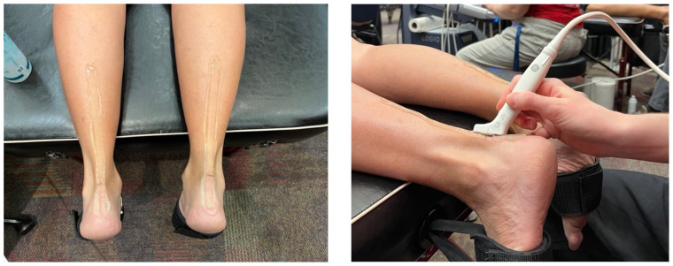
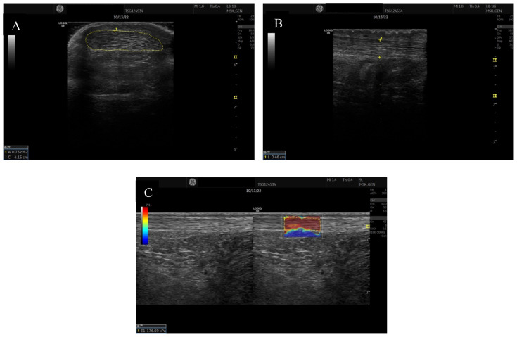
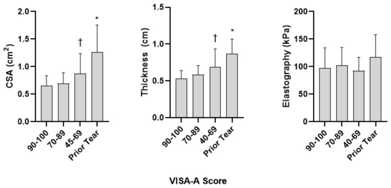
Morphologic Achilles tendon properties obtained via diagnostic ultrasound imaging are valuable in understanding Achilles tendon health and injury. Currently, limited information exists regarding Achilles tendon morphological properties amongst active aging adults based upon Victorian Institute of Sport Assessment (VISA-A) scores. Achilles tendon morphologic properties defined by VISA-A score groupings allow clinicians and researchers to compare data values amongst current patients. Purpose: Comparison of physically active aging adults Achilles tendon morphological properties with various VISA-A scores or a previous Achilles tendon rupture. A convenience sample of 121 participants (71 females, 50 males) at least moderately active and 50 years old, were recruited. Participants completed a VISA-A survey, and assigned groups by scores (Group 1: 90-100, Group 2: 70-89, Group 3: 45-69, Group 4: Previous Achilles tendon tear). Achilles tendon ultrasound imaging occurred at the malleolar line (The apex of the medial and lateral malleolus). Following imaging Achilles tendon cross-sectional area (CSA), thickness, and elastography were measured and analyzed. Participants with a previous Achilles tendon rupture displayed significantly larger tendon CSA and thickness compared with other groups (p<0.05). Individuals with VISA-A scores from 45-69 displayed significantly larger tendon CSA and thickness than participants with scores greater than 90 (p<0.03). No significant differences were noted for elastography between groups (p>0.05). Achilles tendon morphological differences exist based upon pain level in physically active aging adults. Diagnostic ultrasound may be used during assessment and rehabilitation of injured tendon tissue to inform about current tendon tissue properties.
Keywords: Achilles tendinopathy; Diagnostic ultrasound; imaging evaluation.
Figures
Similar articles
-
Evidence-Based High-Loading Tendon Exercise for 12 Weeks Leads to Increased Tendon Stiffness and Cross-Sectional Area in Achilles Tendinopathy: A Controlled Clinical Trial.Sports Med Open. 2022 Dec 20;8(1):149. doi: 10.1186/s40798-022-00545-5. Sports Med Open. 2022. PMID: 36538166 Free PMC article.
-
Eccentric and Isometric Exercises in Achilles Tendinopathy Evaluated by the VISA-A Score and Shear Wave Elastography.Sports Health. 2020 Jul/Aug;12(4):373-381. doi: 10.1177/1941738119893996. Epub 2020 Jan 31. Sports Health. 2020. PMID: 32003647 Free PMC article. Clinical Trial.
-
Differences at the Achilles Insertion Between Adults with Insertional and Midportion Achilles Tendinopathy as Observed Using Ultrasound.Muscles Ligaments Tendons J. 2022 Apr-Jun;12(2):115-121. doi: 10.32098/mltj.02.2022.04. Muscles Ligaments Tendons J. 2022. PMID: 36247413 Free PMC article.
-
Is Platelet-rich Plasma Injection Effective for Chronic Achilles Tendinopathy? A Meta-analysis.Clin Orthop Relat Res. 2018 Aug;476(8):1633-1641. doi: 10.1007/s11999.0000000000000258. Clin Orthop Relat Res. 2018. PMID: 29601383 Free PMC article.
-
Longitudinal microvascularity in Achilles tendinopathy (power Doppler ultrasound, magnetic resonance imaging time-intensity curves and the Victorian Institute of Sport Assessment-Achilles questionnaire): a pilot study.Skeletal Radiol. 2010 Jun;39(6):509-21. doi: 10.1007/s00256-009-0772-0. Epub 2009 Aug 27. Skeletal Radiol. 2010. PMID: 19711073 Review.
References
LinkOut - more resources
Full Text Sources
Miscellaneous