

Epidemiological and Histopathological Features of Oral Squamous Cell Carcinoma-A Retrospective Study
- PMID: 39574818
- PMCID: PMC11578359
- DOI: 10.12865/CHSJ.50.03.08
Epidemiological and Histopathological Features of Oral Squamous Cell Carcinoma-A Retrospective Study
Abstract
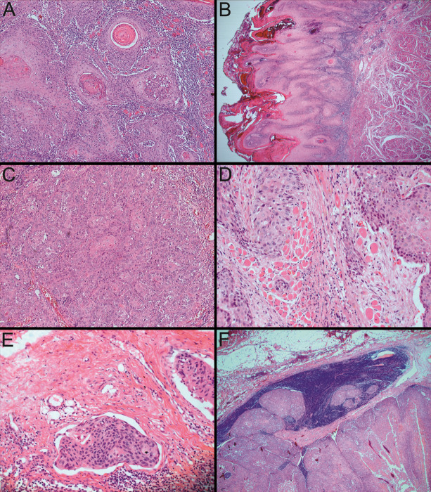
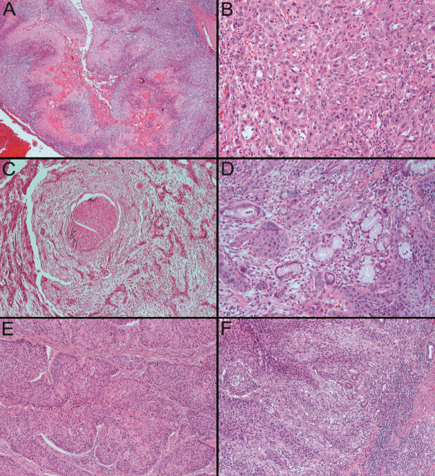
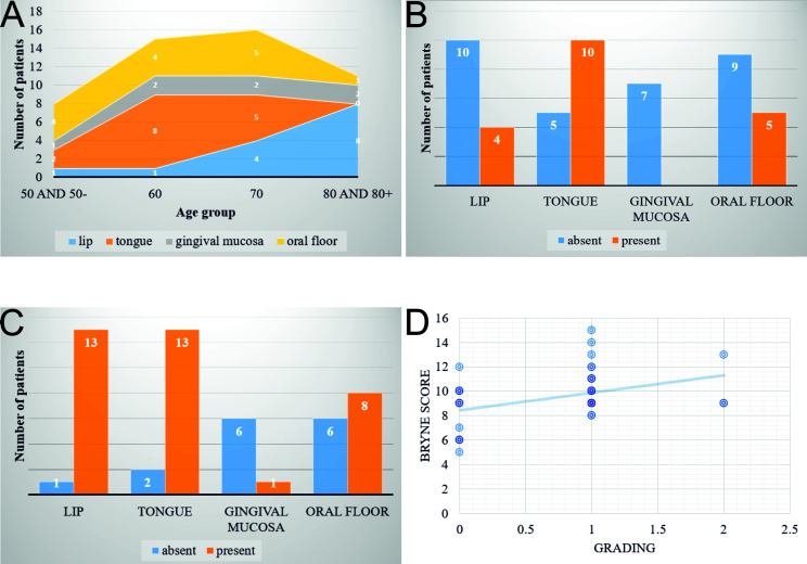
Oral Squamous Cell Carcinoma (OSCC) it was reported to be the 6th on the list of human malignant neoplasms responsible for high morbidity and mortality worldwide. We conducted a retrospective study between 2009-2019, investigating 50 such cancers hospitalized and diagnosed during this period in our institution. The purpose of the study was to establish a clinical-morphological profile of this type of cancer developed in the geographical area served by our institution. The epidemiological study highlighted the predominance of cases in men over 50 years old, mainly affecting the tongue, followed by the lips and oral floor. The histopathological study showed the prevalence of conventional cases of OSCC (70%) and the rest of the cases belonging to rarer forms (acantholytic-18%, verrucous-6%, basaloid-4% and sarcomatoid-2%). In terms of the degree of differentiation, the moderately differentiated cases prevailed (64%) and according to the TNM clinical stage, most cases were diagnosed in stage II (36%) and IV (26%). 70% of investigated cases presented muscle invasion and 38% perineural invasion. Our investigation highlighted the existence of particular morpho-clinical profiles depending on the tumor topography. Thus, tumors developed at the tongue level reached the maximum frequency in the 6th decade of life, being absent in the 8th decade and most often associated muscle invasion and perineural invasion, being diagnosed in advanced pTNM stages.
Keywords: Epidemiology; Histopathology; Oral cavity; Oral squamous cell carcinomas.
Copyright © 2022, Medical University Publishing House Craiova.
Conflict of interest statement
The authors have no conflict of interest to declare.
Figures
References
-
- Capote-Moreno A, Brabyn P, Munoz-Guerra MF, Sastre-Perez J, Escorial-Hernandez V, Rodriguez-Campo FJ, Garcia T, Naval-Gias L. Oral squamous cell carcinoma: epidemiological study and risk factor assessment based on a 39-year series. Int. J. Oral Maxillofac. Surg. 2020;49:1525–1534. - PubMed
-
- Christina McC, Alex K, Marco AM, Iona TL, Tanya J, Grace B. Oral Squamous Cell Carcinoma Associated with Precursor Lesions. Cancer Prev Res. 2021;14(9):873–883. - PubMed
-
- Tiwana MS, Wu J, Hay J, Wong F, Cheung W, Olson RA. year survival outcomes for squamous cell carcinomas of the head and neck: population-based outcomes from a Canadian province. Oral Oncol. 2014;50:651–656. - PubMed
-
- Johnson N , Franceschi S , et al. In: World Health Organization classification of tumours: pathology and genetics of head and neck tumours . Barnes L , Eveson JW , et al., editors. Lyon : IARC ; 2005 . Squamous cell carcinoma ; pp. 168 – 175 .
LinkOut - more resources
Full Text Sources