[Preprint]. 2024 Oct 28:2024.10.26.24316190.
doi: 10.1101/2024.10.26.24316190.

Severe motor impairment is associated with lower contralesional brain age in chronic stroke

Affiliations

Severe motor impairment is associated with lower contralesional brain age in chronic stroke

Gilsoon Park et al. medRxiv. .

Abstract

Background: Stroke leads to complex chronic structural and functional brain changes that specifically affect motor outcomes. The brain-predicted age difference (brain-PAD) has emerged as a sensitive biomarker. Our previous study showed higher global brain-PAD associated with poorer motor function post-stroke. However, the relationship between local stroke lesion load, regional brain age, and motor impairment remains unclear.

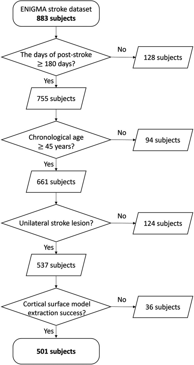
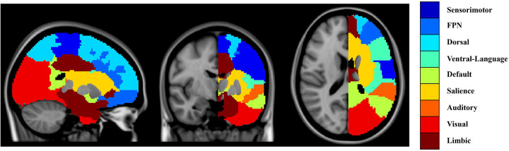
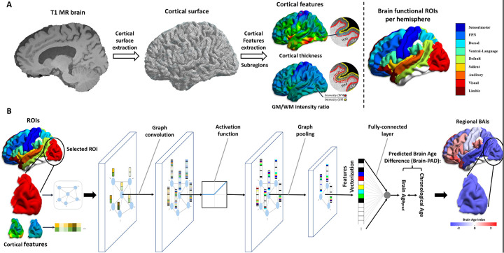
Methods: We studied 501 individuals with chronic unilateral stroke (>180 days post-stroke) from the ENIGMA Stroke Recovery Working Group dataset (34 cohorts). Structural T1-weighted MRI scans were used to estimate regional brain-PAD in 18 predefined functional subregions via a graph convolutional network algorithm. Lesion load for each region was calculated based on lesion overlap. Linear mixed-effects models assessed associations between lesion size, local lesion load, and regional brain-PAD. Machine learning classifiers predicted motor outcomes using lesion loads and regional brain-PADs. Structural equation modeling examined directional relationships among corticospinal tract lesion load (CST-LL), ipsilesional brain-PAD, motor outcomes, and contralesional brain-PAD.

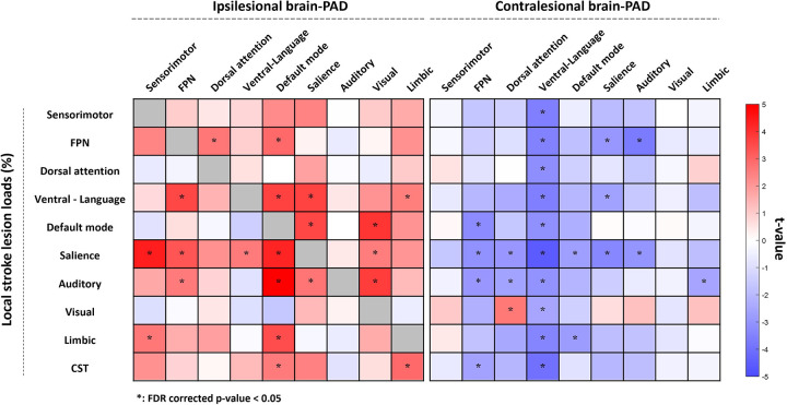
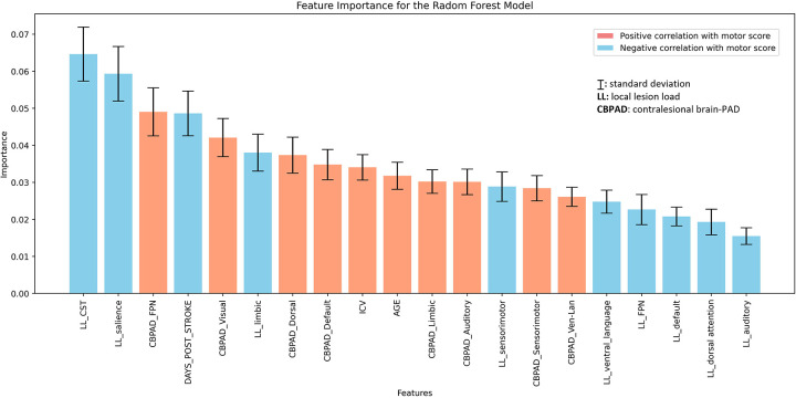
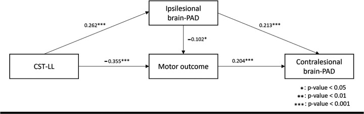
Findings: Larger total lesion size was positively associated with higher ipsilesional regional brain-PADs (older brain age) across most regions (p < 0.05), and with lower contralesional brain-PAD, notably in the ventral attention-language network (p < 0.05). Higher local lesion loads showed similar patterns. Specifically, lesion load in the salience network significantly influenced regional brain-PADs across both hemispheres. Machine learning models identified CST-LL, salience network lesion load, and regional brain-PAD in the contralesional frontoparietal network as the top three predictors of motor outcomes. Structural equation modeling revealed that larger stroke damage was associated with poorer motor outcomes (β = -0.355, p < 0.001), which were further linked to younger contralesional brain age (β = 0.204, p < 0.001), suggesting that severe motor impairment is linked to compensatory decreases in contralesional brain age.

Interpretation: Our findings reveal that larger stroke lesions are associated with accelerated aging in the ipsilesional hemisphere and paradoxically decelerated brain aging in the contralesional hemisphere, suggesting compensatory neural mechanisms. Assessing regional brain age may serve as a biomarker for neuroplasticity and inform targeted interventions to enhance motor recovery after stroke.

Fundings: Micheal J Fox Foundation, National Institutes of Health, Canadian Institutes of Health Research, National Health and Medical Research Council, Australian Brain Foundation, Wicking Trust, Collie Trust, and Sidney and Fiona Myer Family Foundation, National Heart Foundation, Hospital Israelita Albert Einstein, Australian Research Council Future Fellowship, Wellcome Trust, National Institute for Health Research Imperial Biomedical Research Centre, European Research Council, Deutsche Forschungsgemeinschaft, REACT Pilot, National Resource Center, Research Council of Norway, South-Eastern Norway Regional Health Authority, Norwegian Extra Foundation for Health and Rehabilitation, Sunnaas Rehabilitation Hospital HT, University of Oslo, and VA Rehabilitation Research and Development.

Keywords: graph convolutional network; lesion load; motor impairment; regional brain age; stroke outcome prediction; stroke recovery.


Conflict of interest statement

Competing Interests S. C. Cramer is a consultant for Constant Therapeutics, BrainQ, Myomo, MicroTransponder, Panaxium, Beren Therapeutics, Medtronic, Stream Biomedical, NeuroTrauma Sciences, and TRCare; C. A. Hanlon served as a consultant to MagStim, Roswell Park Cancer Insitute, and is an employee of BrainsWay; G. F. Wittenberg serves on the medical advisory boards for Myomo and NeuroInnovators; The other authors report no disclosures relevant to the manuscript.

Figures

Figure 1.
Figure 1.. The data selection flowchart for stroke subjects.
From the 883 initial subjects, we selected 501 participants who met the following four criteria: (1) post-stroke duration of at least 180 days, (2) chronological age of 45 years or older, (3) presence of stroke lesion on only one side of the hemisphere (i.e., unilateral lesion), and (4) successful extraction of cortical thickness.
Figure 2.
Figure 2.. Functional subregions used to define local stroke lesion load.
We extended the atlas defined by Yeo et al. (2011) to include white matter regions. Each voxel in the white matter region was labeled based on a k-nearest neighbor. The subregions of interest include the sensorimotor, frontoparietal, dorsal attention, ventral attention with language, default mode, salience, auditory, visual, and limbic networks, and these were further divided into left and right hemispheres to produce 18 regions of interest (ROIs).
Figure 3.
Figure 3.. The training flowchart for predicting regional brain age.
(A) Flowchart for data generation to pass into graph convolutional networks (GCNs) for predicting regional brain age. We built a cortical surface model from a 3D T1-weighted image using the CIVET pipeline and extracted cortical thickness and gray matter (GM) to white matter (WM) intensity ratio. The cortical surface and cortical features were divided into 18 regions of interest (ROIs). (B) One GCN model per ROI was trained to predict regional brain age. The cortical surface was used to define nodes and edges for a graph structure, and cortical features were used as signals for each node. All models have the same GCN structure, which consists of a graph convolution, rectified linear unit for activation function, max-pooling layer for graph pooling, and a fully connected layer. To obtain regional brain predicted age difference (brain-PAD), we took the difference between the predicted regional brain age and chronological age.
Figure 4.
Figure 4.. Results of the association analysis between lesion loads and regional brain-PAD.
Each square represents the association between regional brain-PAD (x-axis) and lesion load (y-axis). A darker red color indicates higher brain-PAD (older appearing brain), while a darker blue color indicates lower brain-PAD (younger appearing brain). As the brain age in lesional regions (i.e. a lesion load in the ROI for which brain-PAD was computed > 20%) was not computed due to a potentially false brain age prediction, the correlation analyses in the diagonal cells were not performed and colored in gray. The asterisk denotes a significant result (p < 0.05) after False Discovery Rate (FDR) correction. Higher local stroke lesion loads showed correlations with higher ipsilesional and lower contralesional regional brain-PADs. Specifically, ipsilesional regional brain-PADs of FPN, default mode, salience, and visual networks and contralesional regional brain-PADs of FPN, dorsal attention, ventral attention-language, and salience networks were influenced by at least 3 lesion loads. Lesion load in the salience network showed widespread significant influence on both ipsilesional (sensorimotor, FPN, ventral attention-language, default mode, and visual networks) and contralesional regional brain-PAD (FPN, dorsal attention, ventral attention-language, default mode, salience, and auditory networks).
Figure 5.
Figure 5.. Ranking of feature importance scores in predicting motor outcome based on 5,000 iterations bootstrapping of the Random Forest method.
The mean and standard deviation of the feature importance scores are shown for each predictor of motor outcome. The color of the bars indicates whether each feature is significantly correlated with a motor score value, indicating a positive (light red) or negative (light blue) trend. The results highlight the top three features based on mean importance: local lesion load in the corticospinal tract and salience network, and contralesional regional brain-PAD in the FPN. Of the top 20 significant predictors, 8 were contralesional regional brain-PAD and none were ipsilesional regional brain-PAD.
Figure 6.
Figure 6.. Structural equation model (SEM) to determine the relationship between CST-LLs, regional brain-PADs, and motor scores.
This model shows the significant directional relationship between corticospinal tract lesion load (CST-LL), ipsilesional brain-PAD, mean contralesional regional brain-PAD, and motor outcome. The SEM model had acceptable model fit statistics: χ2/df=0.99, χ2 p-value = 0.3717, CFI=1.00, RMSEA=0.00, and AGFI=0.95. All associations in the model were statistically significant (p-value < 0.001) except for the association between ipsilesional brain-PAD and motor outcome. A higher CST-LL correlated with worse motor outcomes (β = −0.355, p-value < 0.001) and higher ipsilateral brain-PAD (β = 0.262, p-value < 0.001). A higher ipsilesional brain-PAD correlated with worse motor outcome (β = −0.102, p-value < 0.05) and higher mean regional brain-PAD in contralesional networks (β = 0.213, p-value < 0.001). A worse motor outcome correlated with lower mean regional brain-PAD of contralesional networks (β = 0.204, p-value < 0.001). We also found that motor outcome mediated the impact of CST-LL on mean regional brain-PAD of contralesional networks. More specifically, CST-LL directly and negatively affected motor outcomes (direct effect=−0.355), with larger lesion load associated with worsened motor impairment. CST-LL indirectly and negatively affected motor outcomes through its effect on ipsilesional brain-PAD (indirect effect=−0.028). On the other hand, CST-LL indirectly and negatively affected mean regional brain-PAD of contralesional networks through its effect on motor outcomes (indirect effect=−0.072). Ipsilesional brain-PAD directly and positively affected the mean regional brain-PAD of contralesional networks (direct effect=0.213).

References

    1. Schlemm E, Schulz R, Bönstrup M, et al. Structural brain networks and functional motor outcome after stroke—a prospective cohort study. Brain Commun. 2020;2(1):fcaa001. doi: 10.1093/braincomms/fcaa001 - DOI - PMC - PubMed
    1. Brodtmann A, Khlif MS, Egorova N, Veldsman M, Bird LJ, Werden E. Dynamic Regional Brain Atrophy Rates in the First Year After Ischemic Stroke. Stroke. 2020;51(9):e183–e192. doi: 10.1161/STROKEAHA.120.030256 - DOI - PubMed
    1. Paul T, Wiemer VM, Hensel L, et al. Interhemispheric Structural Connectivity Underlies Motor Recovery after Stroke. Ann Neurol. 2023;94(4):785–797. doi: 10.1002/ana.26737 - DOI - PubMed
    1. Pinter D, Gattringer T, Fandler-Höfler S, et al. Early Progressive Changes in White Matter Integrity Are Associated with Stroke Recovery. Transl Stroke Res. 2020;11(6):1264–1272. doi: 10.1007/s12975-020-00797-x - DOI - PMC - PubMed
    1. Li W, Li Y, Zhu W, Chen X. Changes in brain functional network connectivity after stroke. Neural Regen Res. 2014;9(1):51–60. doi: 10.4103/1673-5374.125330 - DOI - PMC - PubMed

Publication types

LinkOut - more resources