

Ultrastructural alterations and mitochondrial dysfunction in skeletal muscle of peripheral artery disease patients: implications for early therapeutic interventions
- PMID: 39574966
- PMCID: PMC11579521
- DOI: 10.17179/excli2024-7592
Ultrastructural alterations and mitochondrial dysfunction in skeletal muscle of peripheral artery disease patients: implications for early therapeutic interventions
Abstract
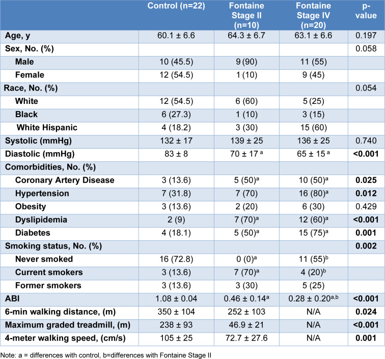
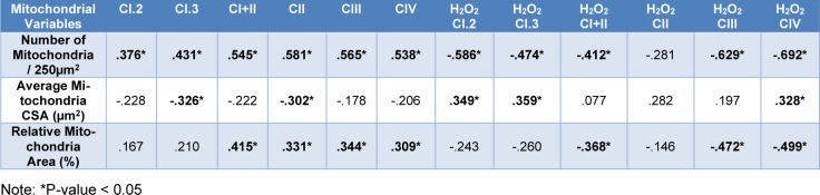
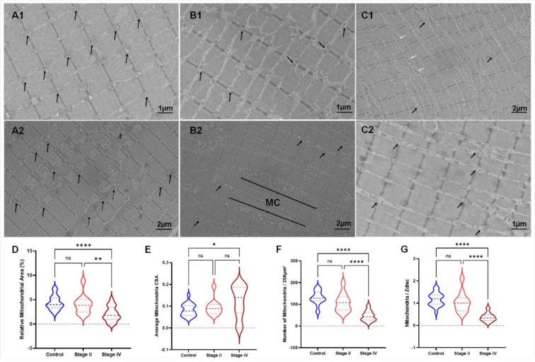
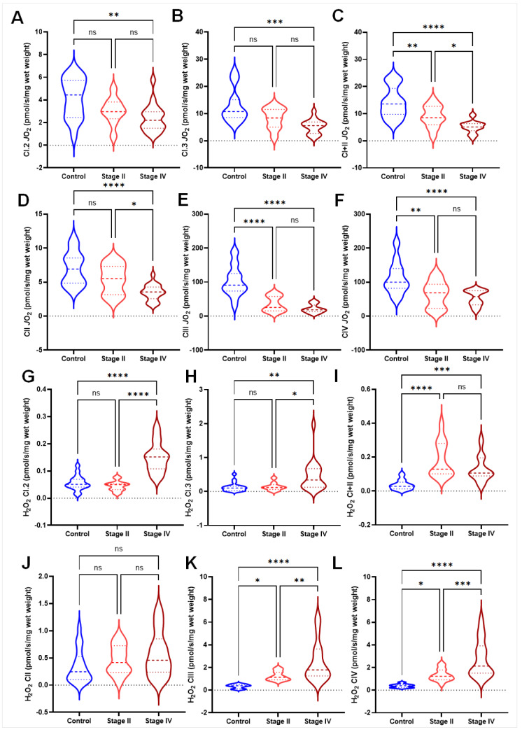
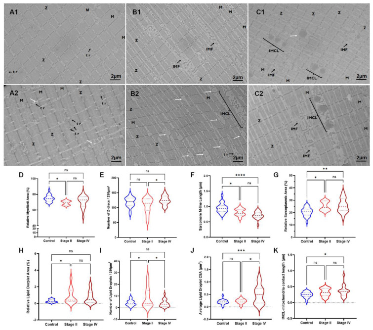
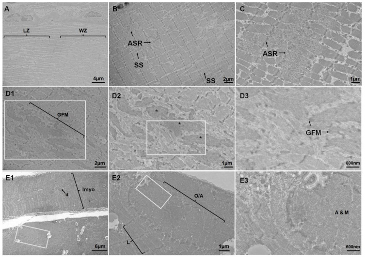
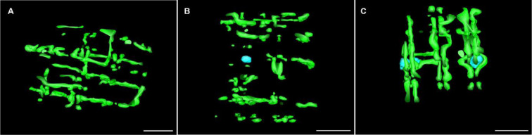
Peripheral artery disease (PAD) is an atherosclerotic condition that impairs blood flow to the lower extremities, resulting in myopathy in affected skeletal muscles. Improving our understanding of PAD and developing novel treatment strategies necessitates a comprehensive examination of cellular structural alterations that occur in the muscles with disease progression. Here we aimed to employ electron microscopy to quantify skeletal muscle ultrastructural alterations responsible for the myopathy of PAD. Fifty-two participants (22 controls, 10 PAD Stage II, and 20 PAD Stage IV) were enrolled. Gastrocnemius biopsies were obtained to determine mitochondrial respiration and oxidative stress. Skeletal muscle sarcomere, mitochondria, lipid droplets, and sarcoplasm were assessed using transmission electron microscopy and focused ion beam scanning electron microscopy. Controls and PAD Stage II patients underwent walking performance tests: 6-minute walking test, 4-minute walking velocity, and maximum graded treadmill test. We identified several prominent ultrastructural modifications in PAD gastrocnemius, including reduced sarcomere dimensions, alterations in mitochondria number and localization, myofibrillar disorientation, changes in lipid droplets, and modifications in mitochondria-lipid droplet contact area. These changes correlated with impaired mitochondrial respiration and increased ROS production. We observed progressive deterioration in mitochondrial parameters across PAD stages. Stage II PAD showed impaired mitochondrial function and structure, while stage IV exhibited further deterioration, more pronounced structural alterations, and a decrease in mitochondrial content. The walking performance of Stage II PAD patients was significantly reduced. Our findings suggest that pathological mitochondria play a key role in the skeletal muscle dysfunction of PAD patients and represent an important target for therapeutic interventions aimed at improving clinical and functional outcomes in this patient population. Our data indicate that treatments should be implemented early and may include therapies designed to preserve and enhance mitochondrial biogenesis and respiration, optimize mitochondrial-lipid droplet interactions, or mitigate oxidative stress. Translational Perspective: Peripheral artery disease (PAD) is characterized by skeletal muscle and mitochondrial dysfunction. Ultrastructural changes in skeletal muscle myofibers and mitochondria morphology can provide significant information on the PAD pathophysiology. Here, we investigated skeletal muscle and mitochondria morphological and functional changes at the sarcomere level and across the disease progression and have found that sarcomere lengths and mitochondria count and function are associated with disease progression, indicating loss of skeletal muscle contractile and metabolic function. Ultrastructural changes in the PAD skeletal muscle can provide significant information in the development of new treatments.
Keywords: intramyocellular lipids; mitochondria; muscle; peripheral artery disease; sarcomere atrophy; sarcoplasm.
Copyright © 2024 Wilburn et al.
Conflict of interest statement
The authors declare no conflict of interest. The funding agencies had no role in the design of the study; in the collection, analyses, or interpretation of data; in the writing of the manuscript, or in the decision to publish the results.
Figures
Similar articles
-
Abnormal accumulation of desmin in gastrocnemius myofibers of patients with peripheral artery disease: associations with altered myofiber morphology and density, mitochondrial dysfunction and impaired limb function.J Histochem Cytochem. 2015 Apr;63(4):256-69. doi: 10.1369/0022155415569348. Epub 2015 Jan 9. J Histochem Cytochem. 2015. PMID: 25575565 Free PMC article.
-
Increased skeletal muscle mitochondrial free radical production in peripheral arterial disease despite preserved mitochondrial respiratory capacity.Exp Physiol. 2018 Jun;103(6):838-850. doi: 10.1113/EP086905. Epub 2018 May 8. Exp Physiol. 2018. PMID: 29604234 Free PMC article.
-
Impaired microcirculatory function, mitochondrial respiration, and oxygen utilization in skeletal muscle of claudicating patients with peripheral artery disease.Am J Physiol Heart Circ Physiol. 2022 May 1;322(5):H867-H879. doi: 10.1152/ajpheart.00690.2021. Epub 2022 Mar 25. Am J Physiol Heart Circ Physiol. 2022. PMID: 35333113 Free PMC article.
-
Skeletal Muscle Pathology in Peripheral Artery Disease: A Brief Review.Arterioscler Thromb Vasc Biol. 2020 Nov;40(11):2577-2585. doi: 10.1161/ATVBAHA.120.313831. Epub 2020 Sep 17. Arterioscler Thromb Vasc Biol. 2020. PMID: 32938218 Free PMC article. Review.
-
Walking Exercise Therapy Effects on Lower Extremity Skeletal Muscle in Peripheral Artery Disease.Circ Res. 2021 Jun 11;128(12):1851-1867. doi: 10.1161/CIRCRESAHA.121.318242. Epub 2021 Jun 10. Circ Res. 2021. PMID: 34110902 Review.
Cited by
-
Multiomic Analysis of Calf Muscle in Peripheral Artery Disease and Chronic Kidney Disease.Circ Res. 2025 Mar 28;136(7):688-703. doi: 10.1161/CIRCRESAHA.124.325642. Epub 2025 Feb 18. Circ Res. 2025. PMID: 39963788 Free PMC article.
-
Targeting Atherosclerosis via NEDD4L Signaling-A Review of the Current Literature.Biology (Basel). 2025 Feb 20;14(3):220. doi: 10.3390/biology14030220. Biology (Basel). 2025. PMID: 40136477 Free PMC article. Review.
References
-
- Bergman BC, Hunerdosse DM, Kerege A, Playdon MC, Perreault L. Localisation and composition of skeletal muscle diacylglycerol predicts insulin resistance in humans. Diabetologia. 2012;55:1140–1150. doi: 10.1007/s00125-011-2419-7. doi: 10.1007/s00125-011-2419-7. Available from: - DOI - DOI - PMC - PubMed
LinkOut - more resources
Full Text Sources